. 2002 Jan 15;22(2):577-83.
doi: 10.1523/JNEUROSCI.22-02-00577.2002.

Prefrontal cortex long-term potentiation, but not long-term depression, is associated with the maintenance of extinction of learned fear in mice

Affiliations

Prefrontal cortex long-term potentiation, but not long-term depression, is associated with the maintenance of extinction of learned fear in mice

Cyril Herry et al. J Neurosci. .

Abstract

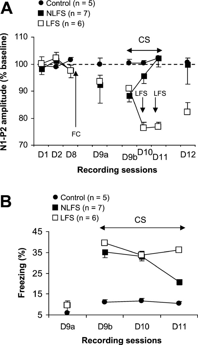
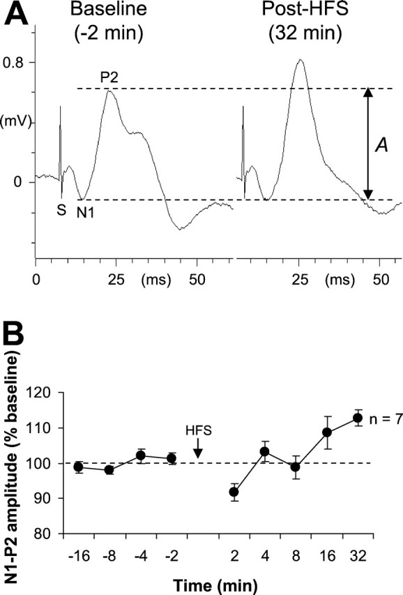
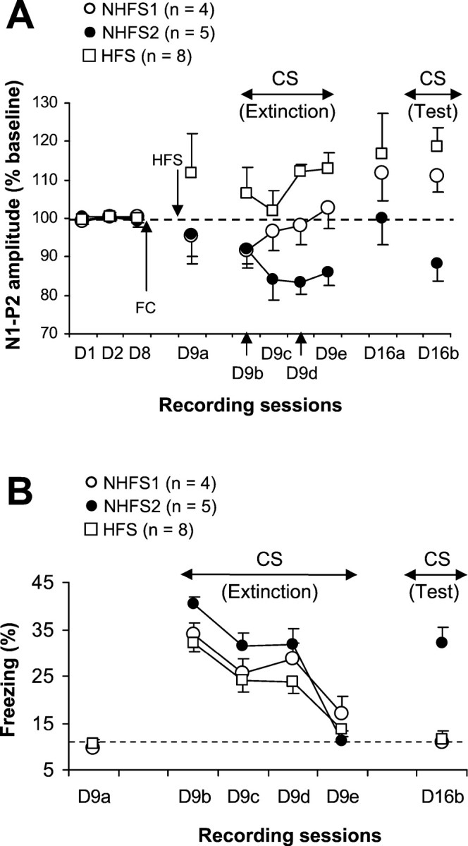
Considerable efforts have been made to identify changes of brain synaptic plasticity associated with fear conditioning. However, for both clinical applications and our fundamental understanding of memory processes, it appears also necessary to investigate synaptic plasticity related to extinction. We previously showed that extinction of freezing to a tone conditioned stimulus (CS; previously paired with footshock) in mice results in a sequence of depression and potentiation of synaptic efficacy in the medial prefrontal cortex (mPFC). These data as well as those from lesion studies suggest that the direction of changes in prefrontal synaptic plasticity may modulate extinction of learned fear. To test this, we analyzed the effects of low-frequency stimulation (LFS) and high-frequency stimulation (HFS) of the mediodorsal thalamic nucleus, known to induce prefrontal long-term depression (LTD) and potentiation (LTP), respectively, on extinction. We found that maintenance of the depression phase, using thalamic LFS, was associated with resistance to extinction. Thalamic HFS applied before extinction testing had no effect on the rate of extinction. However, 1 week follow-up tests revealed that the memory of extinction was intact in these mice (with prefrontal LTP) and in control mice displaying prefrontal LTP-like changes, whereas control mice that did not exhibit such changes displayed a return of freezing to the CS. The results suggest that after extinction the lack of depression-LTP-like conversion sequence in the mPFC synaptic efficacy may profoundly alter the process of consolidation.


Figures

Fig. 1.
Fig. 1.
A, Diagrams of coronal sections of the mouse brain showing electrode placements (dotted areas) in the MD (left) and the mPFC (right). CC, Corpus callosum;HPC, dorsal hippocampus. B, Example of changes in field potential amplitude in a mouse from the LFS group. These representative responses were recorded during the establishment of the baseline (left) and the third sessions of extinction (right). Changes in prefrontal excitability corresponded to changes in the amplitude of the N1–P2 complex (the amplitude A, between the two dotted lines, represents the reference amplitude of the N1–N2 complex).
Fig. 2.
Fig. 2.
A, Mean percentage of changes in N1–P2 amplitude (±SEM) during the 3 d of baseline recording (D1–D8), before the first session (D9a), during sessions (D9b–D11), and 24 hr after the last session (D12) of extinction in conditioned mice that received (LFS group) or did not receive (NLFS group) thalamic LFS and nonconditioned mice (control group). Fear conditioning (FC) took place after the last baseline recording (D8). B, Mean percentage of freezing behavior (±SEM) in the three groups during the 120 sec period preceding the first CS-alone presentation (D9a) and sessions of CS-alone presentations (D9b–D11).
Fig. 3.
Fig. 3.
A, Example of change in MD–mPFC-evoked response after thalamic HFS. These representative responses were recorded during the establishment of the baseline and 32 min after thalamic HFS (the amplitude between the two dotted lines represents the reference amplitude of the N1–P2 complex). B, Mean percentage changes in N1–P2 amplitude (±SEM) during different recording sessions (before and after thalamic HFS).
Fig. 4.
Fig. 4.
A, Mean percentage of changes in N1–P2 amplitude (±SEM) during the 3 d of baseline recording (D1–D8), before the first session (D9a) and during sessions (D9b–D9e) of extinction, before (D16a) and during (D16b) the follow-up test in conditioned mice that received HFS before extinction (HFS group) and their controls that did not receive HFS (NHFS1 and NHFS2 groups). Fear conditioning (FC) took place after the last baseline recording (D8). B, Mean percentage of freezing behavior (±SEM) in the three groups during the 120 sec period preceding the first CS-alone presentation (D9a), during CS-alone presentations (D9b–D9e), and during the 1 week follow-up test of CS-alone presentations (D16b). The dotted line represents mean level of freezing displayed by nonconditioned mice (experiment 1).

Similar articles

Cited by

References

    1. Blanchard RJ, Blanchard C. Crouching as an index of fear. J Comp Physiol Psychol. 1969;67:370–375. - PubMed
    1. Bremner JD, Staib LH, Kaloupek D, Southwick SM, Sooufer R, Charney DS. Neural correlates of exposure to traumatic pictures and sound in Vietnam combat veterans with and without posttraumatic stress disorder: a positron emission tomography study. Biol Psychiatry. 1999a;45:806–816. - PMC - PubMed
    1. Bremner JD, Narayan M, Staib LH, Southwick SM, McGlashan T, Charney DS. Neural correlates of memories of childhood sexual abuse in women with and without posttraumatic stress disorder. Am J Psychiatry. 1999b;156:1787–1795. - PMC - PubMed
    1. Davis HP, Squire LR. Protein synthesis and memory: a review. Psychol Bull. 1984;96:518–559. - PubMed
    1. Ettedgui E, Bridges M. Posttraumatic stress disorder. Psychiatr Clin North Am. 1985;8:89–103. - PubMed

Publication types

LinkOut - more resources