. 2002 Feb;22(3):835-48.
doi: 10.1128/MCB.22.3.835-848.2002.

Role of the Sin3-histone deacetylase complex in growth regulation by the candidate tumor suppressor p33(ING1)

Affiliations

Role of the Sin3-histone deacetylase complex in growth regulation by the candidate tumor suppressor p33(ING1)

A Kuzmichev et al. Mol Cell Biol. 2002 Feb.

Abstract

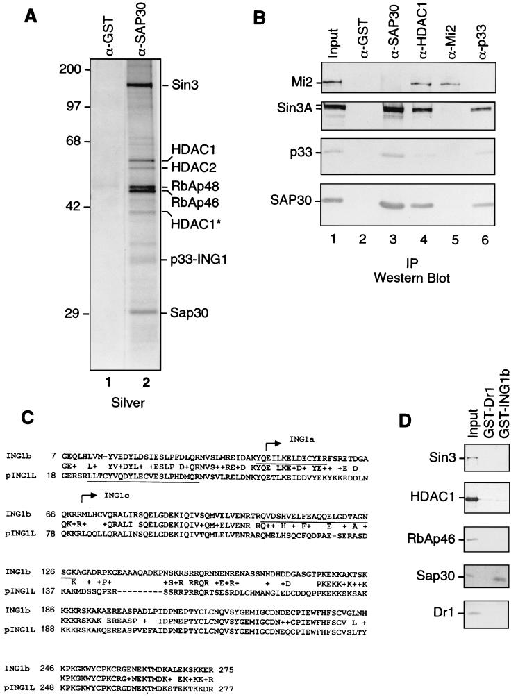
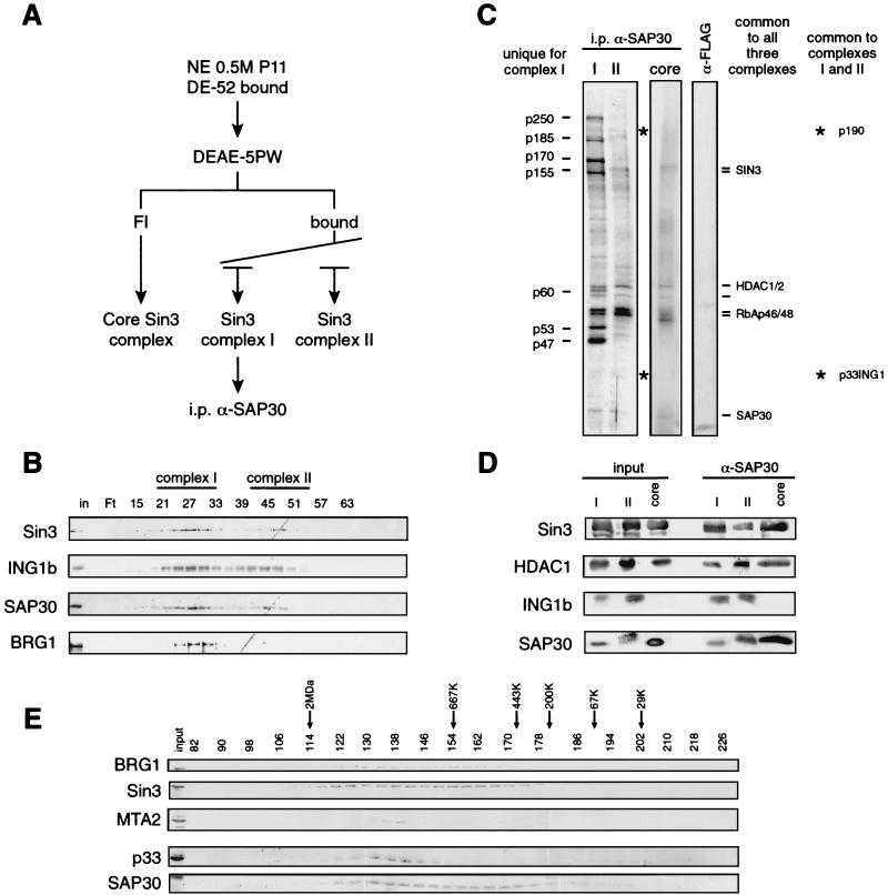
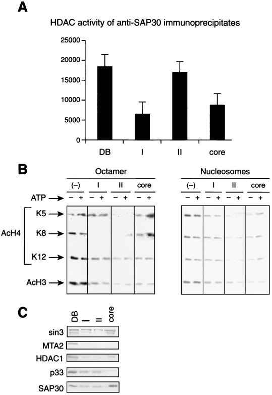
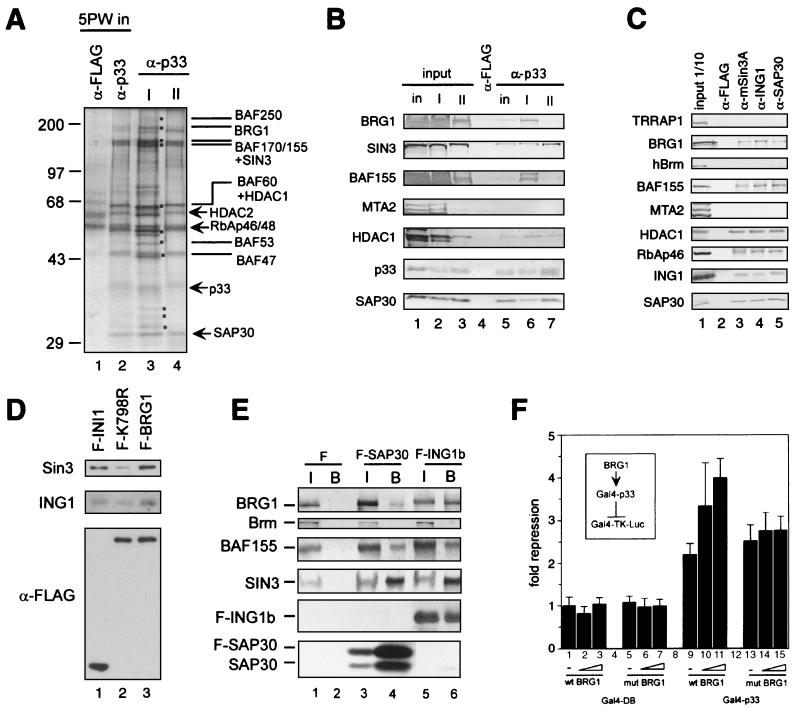
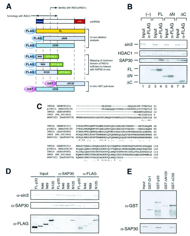
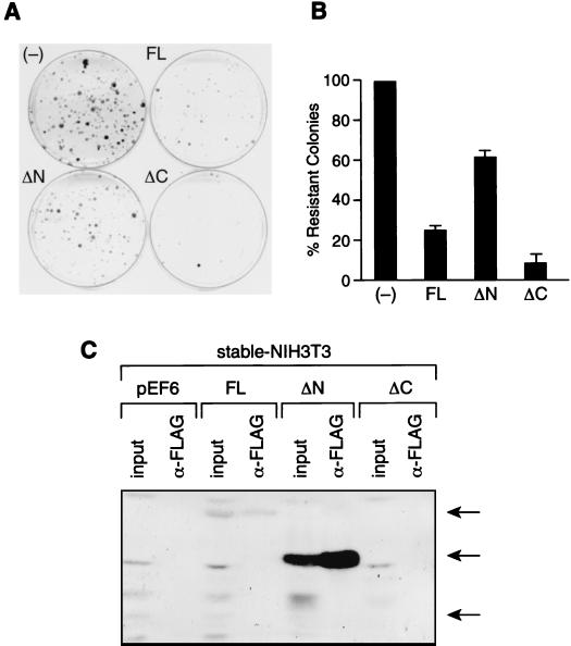
Sin3 is an evolutionarily conserved corepressor that exists in different complexes with the histone deacetylases HDAC1 and HDAC2. Sin3-HDAC complexes are believed to deacetylate nucleosomes in the vicinity of Sin3-regulated promoters, resulting in a repressed chromatin structure. We have previously found that a human Sin3-HDAC complex includes HDAC1 and HDAC2, the histone-binding proteins RbAp46 and RbAp48, and two novel polypeptides SAP30 and SAP18. SAP30 is a specific component of Sin3 complexes since it is absent in other HDAC1/2-containing complexes such as NuRD. SAP30 mediates interactions with different polypeptides providing specificity to Sin3 complexes. We have identified p33ING1b, a negative growth regulator involved in the p53 pathway, as a SAP30-associated protein. Two distinct Sin3-p33ING1b-containing complexes were isolated, one of which associates with the subunits of the Brg1-based Swi/Snf chromatin remodeling complex. The N terminus of p33ING1b, which is divergent among a family of ING1 polypeptides, associates with the Sin3 complex through direct interaction with SAP30. The N-terminal domain of p33 is present in several uncharacterized human proteins. We show that overexpression of p33ING1b suppresses cell growth in a manner dependent on the intact Sin3-HDAC-interacting domain.


Figures

FIG. 1.
FIG. 1.
Identification of p33ING1b as a SAP30-associated protein. (A) Silver staining of anti-SAP30 immunoprecipitates. An aliquot of the DEAE-52 bound material (∼100 μg) was immunoprecipitated with anti-SAP30 antibodies, and immunoprecipitated proteins were washed with buffer containing 0.5 M KCl and 0.05% NP-40. Proteins were eluted with 0.1 M glycine (pH 2.6) and separated by SDS-PAGE followed by silver staining. (B) Western blots of anti-SAP30, anti-HDAC1, anti-Mi2, and anti-p33 immunoprecipitates. Immunoprecipitations (IP) were performed as in panel A. Input, 10 μg of the DEAE-52-bound fraction. Lanes 2 to 6 correspond to 1/10 of the total glycine eluate from the corresponding affinity columns. (C) Sequence alignment of p33ING1b and pING1L. Peptides derived from the 33-kDa band in anti-SAP30 immunoprecipitates are underlined. The sequences of p33ING1b shared with other ING1 isoforms (ING1a and ING1c) are indicated by arrows. (D) Direct interaction between p33 and SAP30. GST-p33 or GST-Dr1 proteins (100 ng) were attached to glutathione-Sepharose beads (5 μl) and incubated with 200 ng of recombinant purified HDAC1, RbAp46, SAP30, Dr1, or in vitro-translated Sin3 protein in buffer containing 0.5 M KCl, 0.1% NP-40, and 0.1 mg of BSA per ml. The bound proteins were washed with the same buffer and eluted with SDS loading buffer. A 1/10 aliquot of material bound to the beads was analyzed by Western blotting as indicated in the figure. Input, 20 ng of recombinant protein.
FIG. 2.
FIG. 2.
p33ING1b is the component of a subset of Sin3-HDAC complexes. (A) Scheme of the immunoaffinity purification of the different SAP30-containing complexes, i.p., immunoprecipitation. (B) Western blot analysis of the DEAE-5PW column fractions. The column was developed as indicated in Materials and Methods. Aliquots of the fractions (5 μl) were separated by SDS-PAGE, and the fractionation of the indicated polypeptides was analyzed by Western blotting using antibodies against Sin3, SAP30, and p33ING1b. Pools used for subsequent anti-SAP30 and anti-p33 immunoprecipitations are indicated (complex I and complex II). (C) Silver staining of anti-SAP30 immunoprecipitates from DEAE-5PW fractions corresponding to the three Sin3 complexes. Equal SAP30 Western blot amounts of DEAE-5PW fractions corresponding to the three Sin3 complexes (I, II, and core; ∼100 μg) were immunoprecipitated with anti-SAP30 antibodies. Bound proteins were washed with buffer containing 0.5 M KCl and 0.05% NP-40 and eluted with 0.1 M glycine (pH 2.6). A 1/10 aliquot of eluted proteins was separated by SDS-PAGE and visualized by silver staining. Polypeptides unique to complex I are indicated on the left side of the panel. Other polypeptides in the three pools are indicated on the right side of the panel. (D) Western blot analysis of anti-SAP30 immunoprecipitates from the DEAE-5PW fractions corresponding to the three Sin3 complexes (I, II, and core). Anti-SAP30 immunoprecipitation was performed as in panel C, and bound proteins were analyzed by Western blotting using antibodies indicated in the figure. Inputs correspond to 1/10 (∼10 μg) of the material used for immunoprecipitation, and the α-SAP30 lanes correspond to 1/10 of the corresponding glycine eluates. (E) Western blot analysis of nuclear extract Sephacryl-400 fractions. Unfractionated HeLa nuclear extract (approximately 10 mg of total protein) was fractionated on a 120-ml Sephacryl-400 column in buffer BC (see Materials and Methods) containing 500 mM KCl, 0.1% NP-40, and 40 μg of ethidium bromide per ml. Aliquots (20 μl) of of each fraction (0.5 ml) were analyzed by Western blotting as indicated.
FIG. 3.
FIG. 3.
HDAC and nucleosome deacetylase activity of the different Sin3 complexes. (A) 3H release assay using core histones. Equal SAP30 Western blot units of each of the Sin3 complexes immunoprecipitated from the DEAE-5PW column fractions using anti-SAP30 antibodies as described in the legend to Fig. 2 (I, II, and core) were incubated with [3H]acetyl-coenzyme A-labeled core histones (0.5 μg, 3 × 10⁷ dpm/μg). Deacetylation activity was measured by quantification of the amount of [3H]acetate released from histones. Anti-SAP30 immunoprecipitate from the DE-52 bound fraction (DB) was used as a positive control. (B) Assay of oligonucleosome deacetylase activity of the native Sin3 complexes I, II, and core, using antibodies specific to particular acetylated lysines. Equal SAP30 Western blot units of partially purified Sin3 complexes were incubated with hyperacetylated core histones or oligonucleosomes (average, five nucleosomes) in the presence or absence of 4 mM ATP, and histone deacetylation was analyzed by Western blotting with antibodies that recognize particular acetylated lysine residues. As a negative control, the same amount of substrate was incubated with a crude fraction devoid of SAP30-HDAC1 (−). (C) Western blot analysis of the partially purified Sin3 complexes (I, II, and core) used for the HDAC assays in panel B. DB corresponds to 5 μg of the DEAE-5PW input. Each lane corresponds to the amount of fraction used for HDAC assays in panel B.
FIG. 4.
FIG. 4.
Interaction between the Sin3 complex I and the Brg1-based Swi-Snf complex. (A) Silver staining of anti-p33 immunoprecipitates derived from the DEAE-5PW fractions corresponding to Sin3 complexes I and II. Equal p33 Western blot units of DEAE-5PW fractions corresponding to Sin3 complexes I and II or DEAE-5PW input (∼100 μg) were immunoprecipitated with anti-p33 or anti-FLAG antibodies, washed with buffer containing 0.5 M KCl and 0.05% NP-40, and eluted with 0.1 M glycine (pH 2.6). Immunoprecipitated proteins (one-quarter of the total glycine eluate) were resolved by SDS-PAGE and visualized by silver staining. Polypeptides specific for complex I are marked by dots. The subunits of the Sin3 and Swi-Snf complexes are indicated. (B) Western blot of anti-p33 immunoprecipitates from DEAE-5PW input and DEAE-5PW fractions corresponding to Sin3 complexes I and II. Anti-p33 immunoprecipitation from DEAE-5PW input (in) and DEAE-5PW fractions corresponding to Sin3 complexes I (I) and II (II) was performed as described in the legend to panel A, followed by Western blot analysis using different antibodies described on the left side of the panel. Input lanes correspond to 10% of the material used for immunoprecipitation. Lanes corresponding to α-p33 immunoprecipitates contain one-quarter of the total glycine eluates. As a negative control, anti-FLAG immunoprecipitation was performed from the DEAE-5PW input material (column labeled α-FLAG). (C) Western blots of anti-Sin3, anti-ING1b, and SAP30 immunoprecipitates from HeLa nuclear extract. Approximately 300 μg of nuclear extracts was immunoprecipitated with antibodies, as indicated at the top of the panel. Samples were washed and proteins were eluted as described in the legend to panel A. Half of the total glycine eluate was separated by SDS-PAGE and analyzed by Western blotting using the antibodies indicated on the left side of the panel. (D) Sin3 and p33ING1b are associated with the Brg1-based Swi-Snf complex. Nuclear extracts from HeLa cells stably expressing FLAG-tagged wild-type Brg1 (F-BRG1) and ATPase-deficient mutant (K798R) (F-K798R) and FLAG-tagged INI1 (F-INI1) were fractionated on an anti-FLAG agarose column, and bound proteins were eluted with excess FLAG peptide and analyzed by Western blotting as described previously (42). To ensure equal efficiency of immunoprecipitation of FLAG-tagged INI1, Brg1, and Brg1(K798R), the same immunoprecipitates were analyzed by Western blotting with anti-FLAG antibodies (bottom panel). Cell lines have been described previously (42, 43). (E) Endogenous Brg1 and BAF155 but not Brm coimmunoprecipitate with transiently overexpressed FLAG-tagged SAP30 and p33ING1b. Nuclear extracts were prepared from 5 × 10⁷ 293T cells transfected with empty vector (F), FLAG-SAP30 (F-SAP30), or FLAG-p33ING1b (F-ING1b) expression vectors. These extracts (300 μg) were immunoprecipitated with anti-FLAG antibodies, and bound proteins were washed with buffer containing 0.5 M KCl and 0.05% NP-40 and eluted with the SDS loading buffer followed by Western blot analysis using antibodies described on the left side of the panel. Input (I) corresponds to 1/10 of the nuclear extract used for immunoprecipitation, whereas the α-FLAG bead-bound fraction (B) corresponds to 1/2 of the material eluted from the beads. (F) Brg1 enhances repression by Gal4-p33ING1b. 293T cells (106 cells per 6-cm plate) were transfected with a luciferase reporter driven by a promoter containing five copies of the Gal4 DNA binding site (Gal-TK-Luc; 250 ng), together with a Gal4-p33 fusion protein (Gal4-p33; 500 ng) or Gal4 DNA-binding domain alone (Gal4-DB; 500 ng) in the presence or absence of the Brg1 expression vector (wtBRG1, or mutBRG1; 250, 500, or 1,000 ng). Luciferase activity was measured as described in Materials and Methods. Fold repression was normalized to reporter activity in the absence of Gal4-p33.
FIG. 5.
FIG. 5.
The unique N-terminal domain of p33ING1b is required for interaction with the Sin3 complex in vivo. (A) Scheme of ING1 deletion mutants. (B) Western blot analysis of anti-FLAG immunoprecipitates from nuclear extracts of NIH 3T3 cells transfected with FLAG-tagged p33ING1b deletion mutants. Nuclear extracts made from 5 × 10⁷ NIH 3T3 cells transfected with empty vector (−), full-length p33 (FL), and p33 deletion mutants (ΔN and ΔC) were immunoprecipitated with anti-FLAG antibodies. Immunoprecipitated proteins were washed with buffer containing 0.5 M KCl and 0.05% NP-40, eluted with SDS loading buffer, and analyzed by Western blotting using antibodies specific for Sin3,HDAC1, and SAP30. Input corresponds to 1/10 of the nuclear extract used for immunoprecipitation, and α-FLAG corresponds to 1/2 of the eluate from the α-FLAG beads. The bottom panel shows expression of the transfected p33ING1b deletion mutants detected using anti FLAG-antibodies. (C) Sequence alignment of proteins containing the domain similar to the N terminus of p33ING1. Alignment was done using the CLUSTALW program at the European Bioinformatics Institute server (http://www2.ebi.ac.uk/clustalw) (48). Asterisks indicate identical positions, colons indicate conserved substitutions, and dots indicates semiconserved (i.e., conserved in most but not all of the aligned sequences) substitutions. (D) Identification of a minimum domain of p33ING1b sufficient to interact with the Sin3 complex in vivo. 239T cells were transfected with either full-length FLAG-tagged p33 protein (FL-p33) or FLAG-tagged fusion proteins containing the N-terminal 46, 69, or 125 amino acids of the p33 protein fused with GFP (N46, N69, and N125). Nuclear extracts from transfected cells (∼1 mg) were immunoprecipitated either with anti-SAP30 or with anti-FLAG antibody, and aliquots of immunoprecipitated proteins (1/2) were analyzed by Western blotting as indicated on the left side of the panel. Inputs correspond to 5 μg (1/200) of nuclear extracts used for immunoprecipitation. (E) The N terminus of p33 is required for the interaction with SAP30 in vitro. GST pull-down was performed as described in the legend to Fig. 1D, using 100 ng of the indicated GST fusion protein and 100 ng of recombinant SAP30. Aliquots of the reactions (1/2) were analyzed by Western blotting using anti-GST and anti-SAP30 antibodies. Input corresponds to 10 ng of recombinant SAP30.
FIG. 6.
FIG. 6.
Interaction of p33ING1b with the Sin3-HDAC complex is important for growth suppression. (A) Colony formation assay with NIH 3T3 cells stably transfected with p33ING1b deletion mutants. NIH 3T3 cells were transfected with empty pEF6 vector (−), pEF6 vector containing full-length p33 (FL), or pEF6 vector containing p33 deletion mutants ΔN and ΔC. Cells expressing pEF6 constructs were selected on blasticidin for 2 weeks, and resistant colonies were stained with Coomassie brilliant blue. (B) Quantification of data in panel A. Blasticidin-resistant colonies were counted manually. Error was determined based on three independent experiments. (C) Analysis of the expression of p33 deletion mutants in transfected cells that survived selection on blasticidin. Nuclear extracts were prepared from equal numbers of blasticidin-resistant cells (mass cultures) that have been stably transfected with empty vector (pEF6) or with pEF6 vector carrying different p33 deletion mutants (FL, ΔN, and ΔC). Aliquots of these extracts (30 μg) were analyzed by Western blotting using anti-FLAG antibodies (columns labeled input). Also, aliquots of these extracts (300 μg) were immunoprecipitated with anti-FLAG antibodies followed by Western blot analysis (columns labeled α-FLAG).

Similar articles

Cited by

References

    1. Alland, L., R. Muhle, H. Hou, Jr., J. Potes, L. Chin, N. Schreiber-Agus, and R. A. DePlnho. 1997. Role for N-CoR and histone deacetylase in Sin3-mediated transcriptional repression. Nature 387:49–55. - PubMed
    1. Altschul, S. F., T. L. Madden, A. A. Schaffer, J. Zhang, Z. Zhang, W. Miller, and D. J. Lipman. 1997. Gapped BLAST and PSI-BLAST: a new generation of protein database search programs. Nucleic Acids Res. 25:3389–3402. - PMC - PubMed
    1. Ausio, J., and K. E. van Holde. 1986. Histone hyperacetylation: its effects on nucleosome conformation and stability. Biochemistry 25:1421–1428. - PubMed
    1. Ayer, D. E., Q. A. Lawrence, and R. N. Eisenman. 1995. Mad-Max transcriptional repression is mediated by temary complex formation with mammalian homologs of yeast repressor Sin3. Cell 80:767–776. - PubMed
    1. Chen, J. D., and R. M. Evans. 1995. A transcriptional co-repressor that interacts with nuclear hormone receptors. Nature 377:454–457. - PubMed

Publication types

MeSH terms

Substances