. 2002 Feb;22(3):874-85.
doi: 10.1128/MCB.22.3.874-885.2002.

Mitotic phosphorylation of histone H3: spatio-temporal regulation by mammalian Aurora kinases

Affiliations

Mitotic phosphorylation of histone H3: spatio-temporal regulation by mammalian Aurora kinases

Claudia Crosio et al. Mol Cell Biol. 2002 Feb.

Abstract

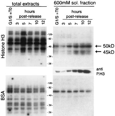
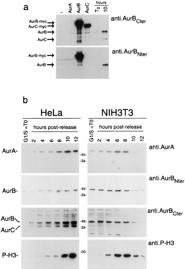
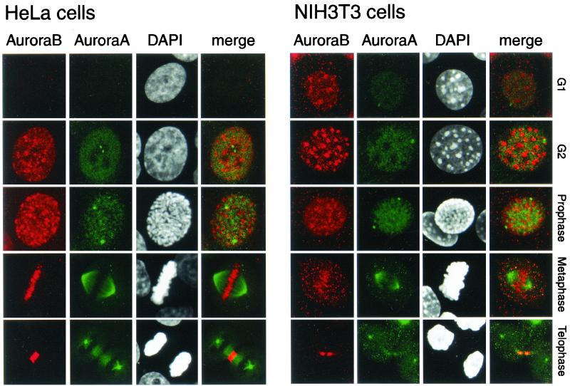
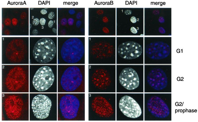
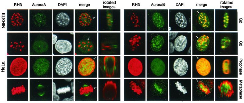
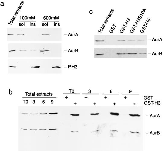
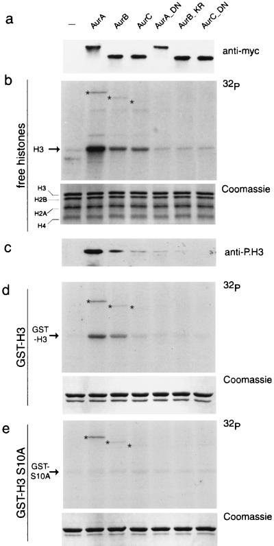
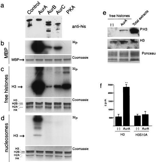
Phosphorylation at a highly conserved serine residue (Ser-10) in the histone H3 tail is considered to be a crucial event for the onset of mitosis. This modification appears early in the G(2) phase within pericentromeric heterochromatin and spreads in an ordered fashion coincident with mitotic chromosome condensation. Mutation of Ser-10 is essential in Tetrahymena, since it results in abnormal chromosome segregation and extensive chromosome loss during mitosis and meiosis, establishing a strong link between signaling and chromosome dynamics. Although mitotic H3 phosphorylation has been long recognized, the transduction routes and the identity of the protein kinases involved have been elusive. Here we show that the expression of Aurora-A and Aurora-B, two kinases of the Aurora/AIK family, is tightly coordinated with H3 phosphorylation during the G(2)/M transition. During the G(2) phase, the Aurora-A kinase is coexpressed while the Aurora-B kinase colocalizes with phosphorylated histone H3. At prophase and metaphase, Aurora-A is highly localized in the centrosomic region and in the spindle poles while Aurora-B is present in the centromeric region concurrent with H3 phosphorylation, to then translocate by cytokinesis to the midbody region. Both Aurora-A and Aurora-B proteins physically interact with the H3 tail and efficiently phosphorylate Ser10 both in vitro and in vivo, even if Aurora-A appears to be a better H3 kinase than Aurora-B. Since Aurora-A and Aurora-B are known to be overexpressed in a variety of human cancers, our findings provide an attractive link between cell transformation, chromatin modifications and a specific kinase system.


Figures

FIG. 1.
FIG. 1.
The activity of two H3 kinases of 50 and 45 kDa correlates with histone H3 mitotic phosphorylation. HeLa cells were synchronized by a double thymidine block and harvested at 0, 3, 5, 7, 10, and 12 h after being released from the block. Progression through the cell cycle and the consequent accumulation of phosphorylated histone H3 was verified by Western blotting using the anti-phosphorylated histone H3 antibody (anti-P.H3). Protein extracts (10 μg) (total extract and 600 mM soluble fractions), prepared at the indicated times, were analyzed by an in-gel kinase assay, using histone H3 (top) or BSA (bottom) as substrates. Equal loading was checked by Coomassie staining.
FIG. 2.
FIG. 2.
Coordinated accumulation of Aurora kinases and H3 phosphorylation during the G2/M transition. (a) Western blot analysis using anti-Aurora-B-Cter and anti-Aurora-B-Nter on 10 μg of total-protein extracts prepared from HeLa cells nontransfected, transfected with myc-tagged Aurora-A, Aurora-B, or Aurora-C, and arrested in G1/S or 10 h after release. (b) Total-protein extracts (10 μg) prepared from synchronized HeLa and NIH 3T3 cells harvested at the indicated times were analyzed by Western blot analysis using anti-Aurora-A, anti-Aurora-B-Cter, anti-Aurora-B-Nter, and anti-P.H3.
FIG. 3.
FIG. 3.
Localization of Aurora-A and Aurora-B in HeLa (left panels) and NIH 3T3 (right panels) cells in G1/S, G2, prophase, metaphase, and telophase. HeLa and NIH 3T3 cells at various stages of the cell cycle were stained for Aurora-B (using anti-Aurora-B-Nter), Aurora-A (using anti-IAK-1), and DNA (using DAPI). Double labeling is indicated as a merge. The cell cycle phases were identified by the DAPI staining.
FIG. 4.
FIG. 4.
Localization of Aurora-A (left panels) and Aurora-B (right panels) in NIH 3T3 cells in G1/S, G2, and G2/prophase. NIH 3T3 cells at various stages of the cell cycle were stained for Aurora-B (using anti-Aurora-B-Nter), Aurora-A (using anti-IAK-1), and DNA (using DAPI). Double labeling is indicated as a merge. The cell cycle phases were identified by the DAPI staining.
FIG. 5.
FIG. 5.
Localization of Aurora-A and Aurora-B in relation to that of P.H3 in HeLa and NIH 3T3 cells during the G2, phase, prophase, and metaphase. NIH 3T3 cells in G2 (top row) and HeLa cells in G2, prophase, and metaphase (bottom three rows) were stained with anti-P.H3 in combination either with anti-IAK-1, to identify Aurora-A (left panels), or with anti-Aurora-BNter, to identify Aurora-B (right panels). The cell cycle phases were identified by the DAPI staining. Double labeling is indicated as a merge. The whole stack of images for each individual panel was subsequentely rotated by 90° on the axis (see arrow) to produce a Z-section (panel “rotated images”).
FIG. 6.
FIG. 6.
Interaction between the N-terminal tail of histone H3 and Aurora-A and Aurora-B. (a) Different susceptibilities of Aurora-A, Aurora-B, and P.H3 to increasing concentrations of salt. Protein extracts were prepared from HeLa cells using buffers with 100 and 600 mM salt concentration and analyzed by Western blotting with anti-IAK-1 to identify Aurora-A (top panel) or with anti-Aurora-B-Nter to identify Aurora-B (bottom panel). (b) Aurora-A and Aurora-B interact with the histone H3 tail. Total-protein extracts from synchronous HeLa cells were incubated with GST alone or GST-H3, and the pulled-down products were tested by Western blotting with anti-Aurora-A (top panel) and anti-Aurora-B-Nter (bottom panel) antibodies. (c) Aurora-A and Aurora-B interact with the histone H4 tail less efficiently than with the H3 tail. Equal amounts of GST fusions (not shown) were verified by Coomassie staining.
FIG. 7.
FIG. 7.
Preferential phosphorylation of H3 at Ser-10 by Aurora kinases. (a) HEK293 cells were transfected with expression plasmids encoding Myc-tagged Aurora-A, Aurora-B, and Aurora-C kinases and the mutated isoforms Aur-A_D274N, Aur-B_K106R, and Aur-C_D184N. Protein extracts prepared from transfected cells were subjected to immunoprecipitation using the anti-Myc antibody. Immunoprecipitated complexes were analyzed by Western blotting to test the expression level of each kinase. (b and c) In vitro kinase assay performed on immunoprecipitated complexes using a histone mix as the substrate. Autoradiography (b, top panel) and Western blotting with anti-P.H3 antibody (c, bottom panel) are shown. The equal amounts of histones in each reaction were verified by Coomassie staining (middle panel). (d and e) In vitro kinase assay performed using the same samples described in panel b and GST-H3 and GSTH3-S10A as substrates; autoradiography (top panel) and Coomassie staining (bottom panel) are shown. The asterisks indicate the bands corresponding to the autophosphorylated Aurora kinases.
FIG. 8.
FIG. 8.
Kinase activity of the recombinant bacterial Aurora kinases. (a) Western blot analysis performed on the recombinant bacterial Aurora kinases using the anti-His antibody. (b to d) In vitro kinase assay performed with recombinant bacterial Aurora-A, Aurora-B, and Aurora-C and protein kinase A on MBP (b), free histones (c), and nucleosomes (d). The presence of equal amounts of histones in each reaction was verified by Coomassie staining. (e) In vitro kinase assay performed with recombinant bacterial Aurora-A analyzed by Western blotting. (f) In vitro kinase assay performed with recombinant bacterial Aurora-A on wild-type and mutated (S10A) histone H3 peptides (aa 1 to 30).

References

    1. Adams, R. R., H. Maiato, W. C. Earnshaw, and M. Carmena. 2001. Essential roles of Drosophila inner centromere protein (INCENP) and aurora B in histone H3 phosphorylation, metaphase chromosome alignment, kinetochore disjunction, and chromosome segregation. J. Cell Biol. 153:865–880. - PMC - PubMed
    1. Ajiro, K., K. Yoda, K. Utsumi, and Y. Nishikawa. 1996. Alteration of cell cycle-dependent histone phosphorylations by okadaic acid. Induction of mitosis-specific H3 phosphorylation and chromatin condensation in mammalian interphase cells. J. Biol. Chem. 271:13197–13201. - PubMed
    1. Bischoff, J. R., L. Anderson, Y. Zhu, K. Mossie, L. Ng, B. Souza, B. Schryver, P. Flanagan, F. Clairvoyant, C. Ginther, C. S. Chan, M. Novotny, D. J. Slamon, and G. D. Plowman. 1998. A homologue of Drosophila aurora kinase is oncogenic and amplified in human colorectal cancers. EMBO J. 17:3052–3065. - PMC - PubMed
    1. Bischoff, J. R., and G. D. Plowman. 1999. The Aurora/Ipl1p kinase family: regulators of chromosome segregation and cytokinesis. Trends Cell Biol. 9:454–459. - PubMed
    1. Bradbury, E. M. 1992. Reversible histone modifications and the chromosome cell cycle. Bioessays 14:9–16. - PubMed

Publication types

MeSH terms