. 2002 Feb;22(3):927-34.
doi: 10.1128/MCB.22.3.927-934.2002.

The Sur7p family defines novel cortical domains in Saccharomyces cerevisiae, affects sphingolipid metabolism, and is involved in sporulation

Affiliations

The Sur7p family defines novel cortical domains in Saccharomyces cerevisiae, affects sphingolipid metabolism, and is involved in sporulation

Michael E Young et al. Mol Cell Biol. 2002 Feb.

Abstract

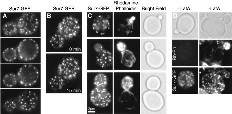
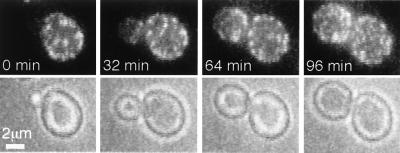
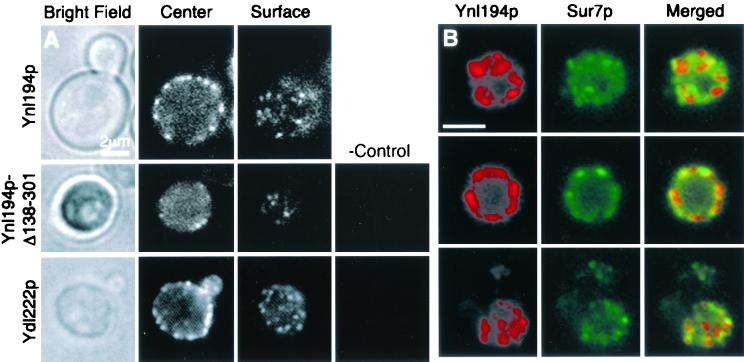
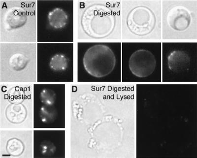
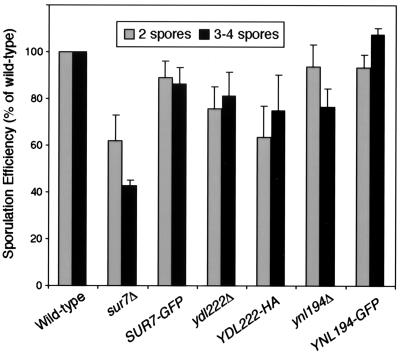
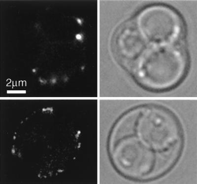
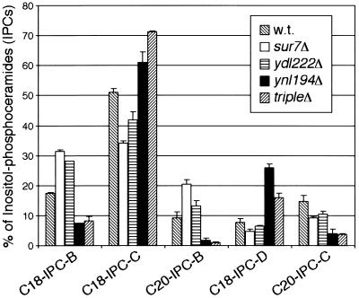
We have discovered a novel cortical patch structure in Saccharomyces cerevisiae defined by a family of integral plasma membrane proteins, including Sur7p, Ynl194p, and Ydl222p. Sur7p-family patches localized as cortical patches that were immobile and stable. These patches were polarized to regions of the cell with a mature cell wall; they were absent from small buds and the tips of many medium-sized buds. These patches were distinct from other known cortical structures. Digestion of the cell wall caused Sur7p patches to disassemble, indicating that Sur7p requires cell wall-dependent extracellular interactions for its localization as patches. sur7Delta, ydl222Delta, and ynl194Delta mutants had reduced sporulation efficiencies. SUR7 was originally described as a multicopy suppressor of rvs167, whose product is an actin patch component. This suppression is probably mediated by sphingolipids, since deletion of SUR7, YDL222, and YNL194 altered the sphingolipid content of the yeast plasma membrane, and other SUR genes suppress rvs167 via effects on sphingolipid synthesis. In particular, the sphingoid base length and number of hydroxyl groups in inositol phosphorylceramides were altered in sur7Delta, ydl222Delta, and yne194Delta strains.


Figures

FIG. 1.
FIG. 1.
Sur7p localizes as cortical, stationary, polarized patches. (A) Fluorescence microscopy of the top, middle, and bottom of a haploid cell containing Sur7-GFP (YJC2054) reveals that Sur7p localizes as patches at the cortex. (B) Time-lapse imaging shows Sur7-GFP patches are stationary and stable. A 15-min interval separates the two single focal plane frames. (C). Fixation and costaining of Sur7-GFP and actin patches, stained with rhodamine-phalloidin, shows that the two signals do not colocalize and that Sur7p patches are not present in small buds. (D) Sur7-GFP patches remain after actin has been depolymerized by latrunculin A (LatA), which was confirmed by rhodamine-phalloidin staining.
FIG. 2.
FIG. 2.
Sur7p patches assemble during bud growth. Selected frames are shown from time-lapse fluorescence microscopy of Sur7-GFP cells (YJC2054). Each frame is a two-dimensional projection of a z-series. The interval between frames is 4 min. The movie is available online (http://www.cooperlab.wustl.edu).
FIG. 3.
FIG. 3.
Ynl194p and Ydl222p localize similarly to Sur7p. All strains were incubated in YPD with 0.4 M NaCl for 75 min prior to viewing to induce YNL194 and YDL222 expression. (A) Single focal planes of Ynl194-GFP (YJC2032) in a living cell show Ynl194-GFP as cortical patches that are absent from the bud (top panels). A Ynl194p construct truncated at the first intracellular residue and tagged with β-galactosidase (YJC2700) localizes similarly to the full-length protein (middle panels). A Ydl222p construct tagged with 3HA before its 14 C-terminal residues (YJC2638) localizes similarly to Sur7p and Ynl194p. The rightmost panels are immunostaining of untagged cells (YJC1193). (B) Cells containing Sur7-GFP and Ynl194p-9myc (YJC2126) were fixed and stained with anti-myc antibodies. Depicted are representative single focal planes showing little (top panels), extensive (middle panels), or partial (bottom panels) colocalization of Sur7p and Ynl194p. Scale bars are 2 μm.
FIG. 4.
FIG. 4.
Disassembly of Sur7p patches upon cell wall digestion. Sur7-GFP cells (YJC2054) were incubated for 15 h at 37°C in the absence (A) or presence (B) of zymolyase and glucuronidase. Cap1-GFP patches (in YJC1423) were retained in the presence of enzymes (C). Sur7-GFP fluorescence was lost from cells that appeared lysed by bright-field microscopy (D).
FIG. 5.
FIG. 5.
Sur7p and its homologues function in sporulation. Strains homozygous for sur7Δ, SUR7-GFP, ydl222Δ, YDL222-3HA, ynl194Δ, or YNL194-GFP (YJC2661, YJC2210, YJC2632, YJC2638, YJC2204, YJC2629) were sporulated on an MSPO plate for 7 days at 30°C. Well-formed asci with three to four clearly visible spores, as well as incomplete asci containing only two spores, were counted. Percentages shown are of the total number of cells and asci. Error bars show the standard error of the mean for 4 to 13 independent experiments, each of which counted 500 to 2,000 cells. Data from each experiment were normalized to results for a wild-type strain (YJC1411) from the same plate, in which 4 to 10% of cells became well-formed asci.
FIG. 6.
FIG. 6.
Sur7p patches during sporulation. A diploid strain homozygous for SUR7-GFP (YJC2210) was sporulated and viewed by fluorescence microscopy. Sur7p patches are visible in the membrane of the ascus, but not the ascospores of mature (top panels) or incomplete (bottom panels) asci. A single focal plane is shown.
FIG. 7.
FIG. 7.
Altered sphingolipid content of sur7Δ, ydl222Δ, and ynl194Δ plasma membranes. Mass spectrometry of plasma membrane lipids from the mutant strains showed differences in the composition of IPC sphingolipids. The IPCs varied in the number of hydroxyl groups (subclasses A to D) and in the length of their sphingoid base component (C18 or C20). Error bars are the standard error of proportion. m/z values are as follows: C18-IPC-B, 936; C18-IPC-C, 952; C20-IPC-B, 964; C18-IPC-D, 968; C20-IPC-C, 980.

References

    1. Amatruda, J. F., and J. A. Cooper. 1992. Purification, characterization and immunofluorescence localization of Saccharomyces cerevisiae capping protein. J. Cell Biol. 117:1067–1076. - PMC - PubMed
    1. Bagnat, M., S. Keränen, A. Shevchenko, and K. Simons. 2000. Lipid rafts function in biosynthetic delivery of proteins to the cell surface in yeast. Proc. Natl. Acad. Sci. USA 97:3254–3259. - PMC - PubMed
    1. Balguerie, A., P. Sivadon, M. Bonneu, and M. Aigle. 1999. Rvs167p, the budding yeast homolog of amphiphysin, colocalizes with actin patches. J. Cell Sci. 112:2529–2537. - PubMed
    1. Barral, Y., V. Mermall, M. S. Mooseker, and M. Snyder. 2000. Compartmentalization of the cell cortex by septins is required for maintenance of cell polarity in yeast. Mol. Cell 5:841–851. - PubMed
    1. Baudin, A., O. Ozierkalogeropoulos, A. Denouel, F. Lacroute, and C. Cullin. 1993. A simple and efficient method for direct gene deletion in Saccharomyces cerevisiae. Nucleic Acids Res. 21:3329–3330. - PMC - PubMed

Publication types

MeSH terms