

RAD1 controls the meiotic expansion of the human HRAS1 minisatellite in Saccharomyces cerevisiae
- PMID: 11784870
- PMCID: PMC133548
- DOI: 10.1128/MCB.22.3.953-964.2002
RAD1 controls the meiotic expansion of the human HRAS1 minisatellite in Saccharomyces cerevisiae
Abstract
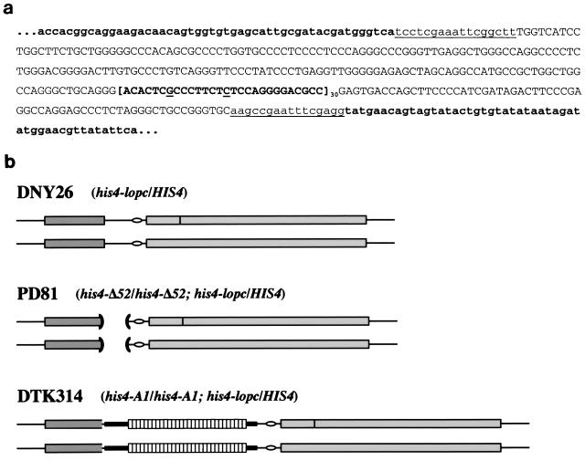
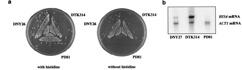
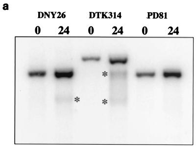
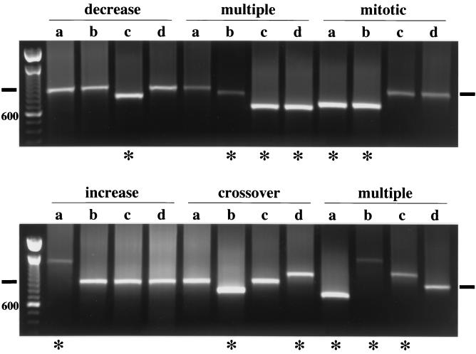
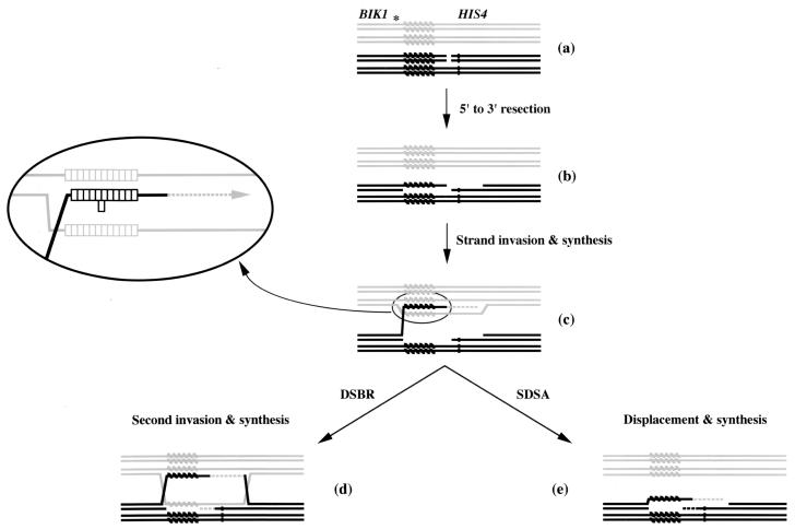
Minisatellite DNA is repetitive DNA with a repeat unit length from 15 to 100 bp. While stable during mitosis, it destabilizes during meiosis, altering both in length and in sequence composition. The basis for this instability is unknown. To investigate the factors controlling minisatellite stability, a minisatellite sequence 3' of the human HRAS1 gene was introduced into the Saccharomyces cerevisiae genome, replacing the wild-type HIS4 promoter. The minisatellite tract exhibited the same phenotypes in yeast that it exhibited in mammalian systems. The insertion stimulated transcription of the HIS4 gene; mRNA production was detected at levels above those seen with the wild-type promoter. The insertion stimulated meiotic recombination and created a hot spot for initiation of double-strand breaks during meiosis in the regions immediately flanking the repetitive DNA. The tract length altered at a high frequency during meiosis, and both expansions and contractions in length were detected. Tract expansion, but not contraction, was controlled by the product of the RAD1 gene. RAD1 is the first gene identified that controls specifically the expansion of minisatellite tracts. A model for tract length alteration based on these results is presented.
Figures
Similar articles
-
The role of CSM3, MRC1, and TOF1 in minisatellite stability and large loop DNA repair during meiosis in yeast.Fungal Genet Biol. 2013 Jan;50:33-43. doi: 10.1016/j.fgb.2012.10.007. Epub 2012 Nov 17. Fungal Genet Biol. 2013. PMID: 23165348 Free PMC article.
-
Length and sequence heterozygosity differentially affect HRAS1 minisatellite stability during meiosis in yeast.Genetics. 2005 Jun;170(2):601-12. doi: 10.1534/genetics.104.026278. Epub 2005 Apr 16. Genetics. 2005. PMID: 15834153 Free PMC article.
-
The influence of sequence divergence between alleles of the human MS205 minisatellite incorporated into the yeast genome on length-mutation rates and lethal recombination events during meiosis.J Mol Biol. 2002 May 31;319(2):315-27. doi: 10.1016/S0022-2836(02)00292-9. J Mol Biol. 2002. PMID: 12051909
-
The multiple roles of the Mre11 complex for meiotic recombination.Chromosome Res. 2007;15(5):551-63. doi: 10.1007/s10577-007-1147-9. Chromosome Res. 2007. PMID: 17674145 Review.
-
[Mechanisms and control of meiotic recombination in the yeast Saccharomyces cerevisiae].J Soc Biol. 1999;193(1):23-7. J Soc Biol. 1999. PMID: 10851552 Review. French.
Cited by
-
The role of CSM3, MRC1, and TOF1 in minisatellite stability and large loop DNA repair during meiosis in yeast.Fungal Genet Biol. 2013 Jan;50:33-43. doi: 10.1016/j.fgb.2012.10.007. Epub 2012 Nov 17. Fungal Genet Biol. 2013. PMID: 23165348 Free PMC article.
-
Stimulation of gross chromosomal rearrangements by the human CEB1 and CEB25 minisatellites in Saccharomyces cerevisiae depends on G-quadruplexes or Cdc13.PLoS Genet. 2012;8(11):e1003033. doi: 10.1371/journal.pgen.1003033. Epub 2012 Nov 1. PLoS Genet. 2012. PMID: 23133402 Free PMC article.
-
The yeast Pif1 helicase prevents genomic instability caused by G-quadruplex-forming CEB1 sequences in vivo.PLoS Genet. 2009 May;5(5):e1000475. doi: 10.1371/journal.pgen.1000475. Epub 2009 May 8. PLoS Genet. 2009. PMID: 19424434 Free PMC article.
-
A Whole Genome Screen for Minisatellite Stability Genes in Stationary-Phase Yeast Cells.G3 (Bethesda). 2013 Apr 9;3(4):741-756. doi: 10.1534/g3.112.005397. G3 (Bethesda). 2013. PMID: 23550123 Free PMC article.
-
Comparative genomics and molecular dynamics of DNA repeats in eukaryotes.Microbiol Mol Biol Rev. 2008 Dec;72(4):686-727. doi: 10.1128/MMBR.00011-08. Microbiol Mol Biol Rev. 2008. PMID: 19052325 Free PMC article. Review.
References
-
- Alani, E., R. Padmore, and N. Kleckner. 1990. Analysis of wild-type and rad50 mutants of yeast suggests an intimate relationship between meiotic chromosome synapsis and recombination. Cell 61:419–436. - PubMed
-
- Allers, T., and M. Lichten. 2001. Differential timing and control of noncrossover and crossover recombination during meiosis. Cell 106:47–57. - PubMed
-
- Applegren, H., H. Cederberg, and U. Rannug. 1999. Meiotic interallelic conversion at the human minisatellite MS32 in yeast triggers recombination in several chromatids. Gene 239:29–38. - PubMed
-
- Applegren, H., H. Cederberg, and U. Rannug. 1997. Mutations at the human minisatellite MS32 integrated in yeast occur with high frequency in meiosis and involve complex recombination events. Mol. Gen. Genet. 256:7–17. - PubMed
Publication types
MeSH terms
Substances
LinkOut - more resources
Full Text Sources
Molecular Biology Databases
Research Materials
Miscellaneous