. 2002 Jan;160(1):101-12.
doi: 10.1016/s0002-9440(10)64354-4.

CD36, a class B scavenger receptor, is expressed on microglia in Alzheimer's disease brains and can mediate production of reactive oxygen species in response to beta-amyloid fibrils

Affiliations

CD36, a class B scavenger receptor, is expressed on microglia in Alzheimer's disease brains and can mediate production of reactive oxygen species in response to beta-amyloid fibrils

Indra Sethy Coraci et al. Am J Pathol. 2002 Jan.

Abstract

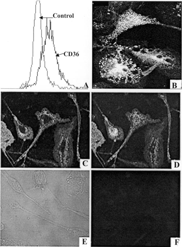
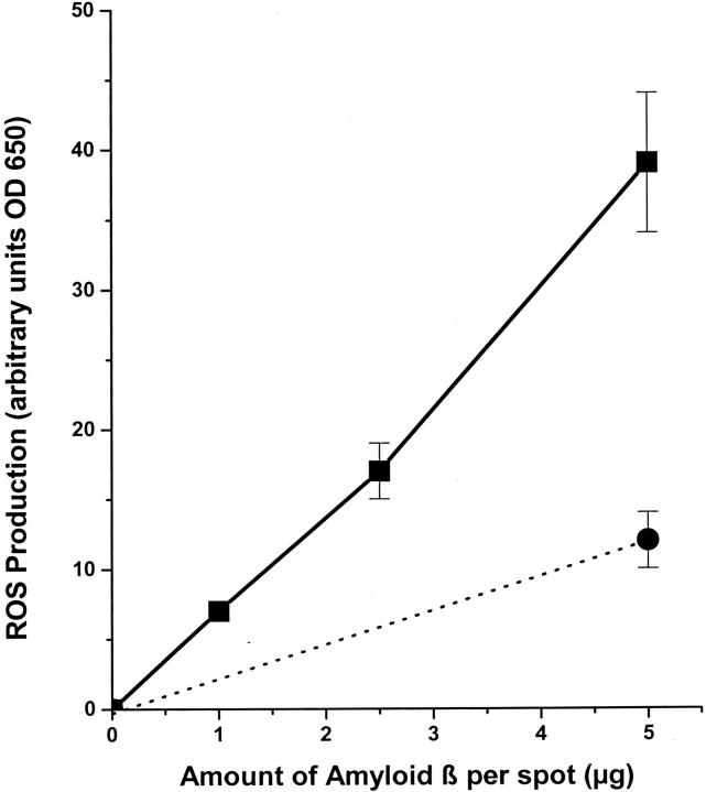
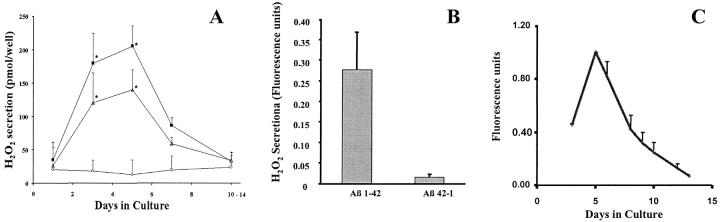
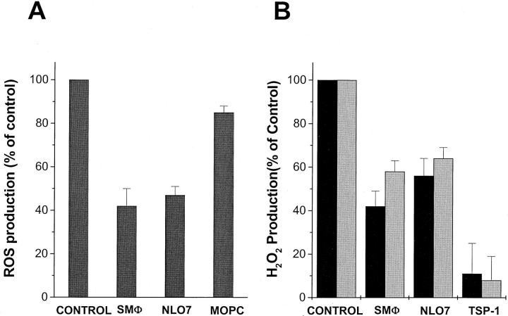
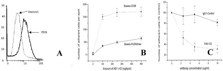
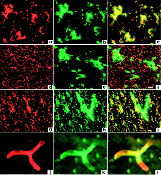
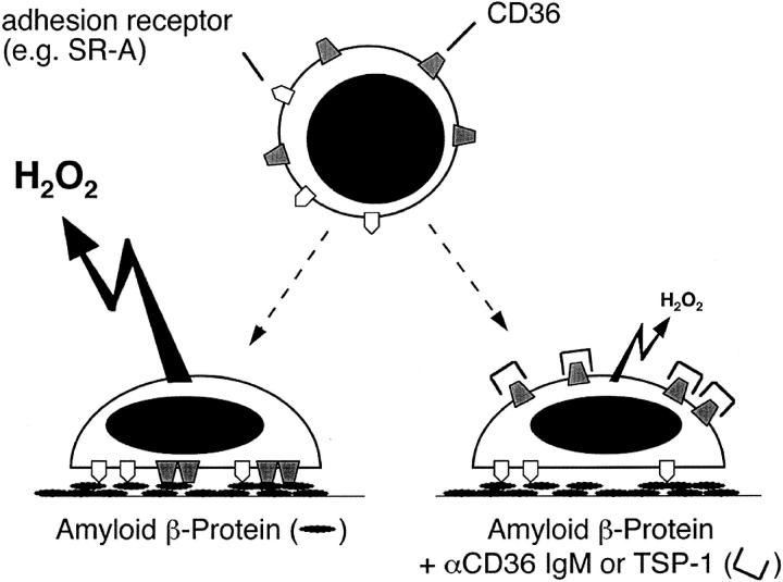
A pathological hallmark of Alzheimer's disease is the senile plaque, composed of beta-amyloid fibrils, microglia, astrocytes, and dystrophic neurites. We reported previously that class A scavenger receptors mediate adhesion of microglia and macrophages to beta-amyloid fibrils and oxidized low-density lipoprotein (oxLDL)-coated surfaces. We also showed that CD36, a class B scavenger receptor and an oxLDL receptor, promotes H(2)O(2) secretion by macrophages adherent to oxLDL-coated surfaces. Whether CD36 is expressed on microglia, and whether it plays a role in secretion of H(2)O(2) by microglia interacting with fibrillar beta-amyloid is not known. Using fluorescence-activated cell sorting analysis and immunohistochemistry, we found that CD36 is expressed on human fetal microglia, and N9-immortalized mouse microglia. We also found that CD36 is expressed on microglia and on vascular endothelial cells in the brains of Alzheimer's disease patients. Bowes human melanoma cells, which normally do not express CD36, gained the ability to specifically bind to surfaces coated with fibrillar beta-amyloid when transfected with a cDNA encoding human CD36, suggesting that CD36 is a receptor for fibrillar beta-amyloid. Furthermore, two different monoclonal antibodies to CD36 inhibited H(2)O(2) production by N9 microglia and human macrophages adherent to fibrillar beta-amyloid by approximately 50%. Our data identify a role for CD36 in fibrillar beta-amyloid-induced H(2)O(2) production by microglia, and imply that CD36 can mediate binding to fibrillar beta-amyloid. We propose that similar to their role in the interaction of macrophages with oxLDL, class A scavenger receptors and CD36 play complimentary roles in the interactions of microglia with fibrillar beta-amyloid.


Figures

Figure 1.
Figure 1.
Characterization of Aβ peptides. A and B: Solutions containing Aβ 42-1 and 1-42 were prepared as described in Materials and Methods and used to coat multispot slides. A: Spots coated with Aβ 42-1 and incubated with Thioflavin S for 1 minute remained unstained implying no fibril formation. B: Spots coated with Aβ 1-42 and incubated with Thioflavin S (Sigma) for 1 minute stained intensely. C: A transmission electron micrograph of negatively stained Aβ 1-42 fibrils (original magnification, ×65,000). D: Nonreducing SDS-PAGE of fAβ 1-42 and 42-1 peptides stained with Coomassie blue (Sigma). The lane containing fAβ 1-42 peptides shows two bands, one at ≅4.2 kd and one at ≅21 kd, whereas the lane containing 42-1 contains a single band at ≅4.2 kd.
Figure 2.
Figure 2.
N9 mouse microglia and cultured human fetal microglia express CD36. A: N9 microglia were incubated with phycoerythrin (PE)-labeled monoclonal anti-CD36 antibody clone SMφ (PE-SMφ) or control antibody (PE-MOPC-104E). Cell-associated fluorescence was measured using FACS analysis as described in Materials and Methods. Mean fluorescence was 197.2 for PE-SMφ-labeled cells and 53.9 for PE-MOPC-labeled cells. B: Adherent human fetal microglia were incubated with PBS containing 1 mg/ml albumin and 5 μg/ml DiI-AcLDL for 4 hours at 37°C, washed, fixed in 3.7% formalin, and visualized using a Zeiss fluorescence microscope. C and D: Adherent human fetal microglia stained with the anti-CD36 monoclonal antibody FA6-152 as described in Materials and Methods. Surface staining was visualized by confocal microscopy of cells for a single optical section (C) and for the entire projection series of 16 sections (D). Cells did not stain with an isotype-matched control antibody (E and F).
Figure 3.
Figure 3.
Fibrillar Aβ 1-42 stimulates cultured human fetal microglia to produce ROS. Human fetal microglia were incubated on multispot slides containing 1, 2.5, or 5 μg of fibrillar Aβ 1-42 (filled squares) or 5 μg of reverse (42-1) nonfibrillar amyloid (filled circle). The amount of ROS produced was measured by NBT reduction as described in Materials and Methods.
Figure 4.
Figure 4.
The capacity of monocyte-derived macrophages to produce H2O2 parallels their expression of CD36. A: Human monocytes/macrophages, cultured for 1 to 14 days in Teflon beakers, were added to wells coated with 2.5 μg of collagen IV overlayed with 2 μg of fibrillar Aβ 1-42 (open triangle), fibrillar Aβ 25-35 (filled square), or Aβ 35-25 (open circle) and incubated for 3 hours. H2O2 production was determined using the Molecular Probes Hydrogen Peroxide Assay Kit as described in Materials and Methods. Data presented are mean values ± SEM above background (collagen IV alone) for six experiments, each done in triplicate. B: H2O2 production by human monocyte-derived macrophages cultured for 5 days in Teflon beakers. Cells were incubated as in A above in wells containing collagen IV alone or collagen IV and 2 μg of fAβ 1-42 or 2 μg of Aβ 42-1. Data presented are mean values ± SEM above background (collagen IV alone) for three experiments each done in triplicate. *, Indicates values significantly different (P < 0.05) from control (open circles), using Student’s test. C: Summary of FACS analyses of CD36 expression on the surfaces of human monocytes/macrophages maintained in Teflon beakers and assayed at the times indicated as described in Materials and Methods. H2O2 production and CD36 expression were both maximal in cells cultured for 5 days.
Figure 5.
Figure 5.
CD36 mediates ROS production by microglia and H2O2 production by macrophages plated on fAβ-coated surfaces. A: N9 microglia (106/ml) were preincubated for 30 minutes with anti-CD36 monoclonal antibody SMφ or NL07 (20 μg/ml) or isotype-matched control antibody MOPC-104E (20 μg/ml). Cells (50,000) were added to each spot of multispot slides coated with fibrillar Aβ 1-42 (black bars). ROS production was measured using the NBT assay as described in Materials and Methods. B: Human monocyte-derived macrophages (4 to 6 days in culture) were preincubated with anti-CD36 mAbs (SMφ or NL07, 20 μg/ml) or TSP-1 (5 μg/ml), plated on surfaces coated with 2.5 μg of collagen IV and 2 μg of fibrillar Aβ 1-42 (black bars) or fibrillar Aβ 25-35 (gray bars) and assayed for H2O2 secretion as described in Materials and Methods. Data are mean values ± SEM of three experiments done in triplicate. ROS production in response to Aβ 42-1 was similar to background (see Figure 4B ▶ , and data not shown).
Figure 6.
Figure 6.
Bowes cells expressing CD36 gain the ability to bind to fAβ 1-42-coated surfaces. A: FACS analysis of Bowes melanoma cells transfected with a mammalian expression vector for human CD36 and stained with a monoclonal antibody FA6-152 to CD36. B: Adhesion Bowes cells (2.5 × 10 cells/ml), transfected with a mammalian expression vector for CD36 (CDM8-CD36) or with a control vector (PcDNAneo), and suspended in phosphate buffer containing 5 mmol/L EDTA, to multispot slides coated with 2 μg of collagen IV and the indicated amount of Aβ 1-42. C: Bowes melanoma cells expressing CD36 were incubated with the indicated concentrations of anti-CD36 monoclonal antibody FA6-152 or an isotype-matched IgG1 control and plated on multispot slides coated with 2 μg of collagen IV and 500 ng of Aβ 1-42.
Figure 7.
Figure 7.
CD36 is expressed by microglia and endothelial cells in AD brains. Staining of frontal cortex from 13 patients was performed using New England Nuclear’s Tyramide Signal Amplification kit as described under Materials and Methods. Each row represents immunofluorescent staining of a single section from an AD brain and is typical for all sections examined in this study. Staining with anti-CD36 antibody is shown in a, d, g, and j, anti-MAC-1 (CD11b) antibody in b and h, anti-GFAP in e, and staining with Ricin in k. Overlays of staining in each row are shown in c, f, i, and l. Cells lining brain vasculature (likely endothelial cells) (k) express CD36 (j and l) but not the microglial marker Mac-1 (g, h, and i).
Figure 8.
Figure 8.
Cooperation between scavenger receptors and CD36 mediates microglial release of ROS. Diagram summarizing proposed effects of CD36 on ROS production by microglia adherent to surfaces coated with Aβ fibrils. SR-A and other scavenger receptors mediate microglial adhesion to fAβ-coated surfaces. Engagement of CD36 by substrate bound fAβ signals ROS and H2O2 production. Pre-exposure of cells to antibodies against CD36 or the CD36 ligand TSP-1 blocks interactions between CD36 and fAβ resulting in inhibition of ROS and H2O2 production (see Figures 2 and 4 ▶ ▶ ).

Similar articles

Cited by

References

    1. Selkoe DJ: The origins of Alzheimer disease: a is for amyloid. JAMA 2000, 283:1615-1617 - PubMed
    1. Wisniewski HM, Robe A, Zigman W, Silverman W: Neuropathological diagnosis of Alzheimer disease. J Neuropathol Exp Neurol 1989, 48:606-609 - PubMed
    1. El Khoury J, Hickman SE, Thomas CA, Loike JD, Silverstein SC: Microglia, scavenger receptors, and the pathogenesis of Alzheimer’s disease. Neurobiol Aging 1998, 19:S81-S84 - PubMed
    1. Akiyama H: Inflammatory response in Alzheimer’s disease. Tohoku J Exp Med 1994, 174:295-303 - PubMed
    1. Meda L, Cassatella MA, Szendrei GI, Otvos Jr L, Baron P, Villalba M, Ferrari D, Rossi F: Activation of microglial cells by beta-amyloid protein and interferon-gamma. Nature 1995, 374:647–650 - PubMed

Publication types

MeSH terms