. 2002 Jan;160(1):131-9.
doi: 10.1016/S0002-9440(10)64357-X.

Podocin localizes in the kidney to the slit diaphragm area

Affiliations

Podocin localizes in the kidney to the slit diaphragm area

Séverine Roselli et al. Am J Pathol. 2002 Jan.

Abstract

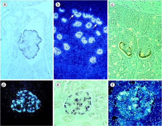
We recently cloned a novel gene, NPHS2, involved in autosomal recessive steroid-resistant nephrotic syndrome. This gene encodes a novel podocyte protein, podocin. Given its similarity with the stomatin family proteins, podocin is predicted to be an integral membrane protein with a single membrane domain forming a hairpin-like structure placing both N- and C-termini in the cytosol. Here, we show by in situ hybridization, that during development, the NPHS2 transcript is first expressed in mesonephric podocytes from the S-shaped body and, later, in the metanephric kidney, in the future podocytes at the late S-shaped body stage. In the mature kidney, NPHS2 is exclusively expressed in the podocytes of mature glomeruli. We generated rabbit polyclonal antibodies against fusion proteins derived from the N- and the C-terminal regions of podocin which detected a single band of 49-kd in transfected HEK293 cell lysates by immunoprecipitation and Western blotting. By immunohistology, podocin was detected in podocytes from the early capillary loop stage in the developing nephrons, and at the basal pole, along the GBM, in mature glomeruli. By electron microscopy, we demonstrate that podocin is facing the slit diaphragm with its two ends in the cytoplasm of the foot processes, in agreement with its predicted structure. Our results suggest that podocin could serve to anchor directly or indirectly components of the slit diaphragm to the cytoskeleton.


Figures

Figure 1.
Figure 1.
Characterization of podocin antibodies by Western blotting and immunoprecipitation. a: Lysates from non-transfected HEK293 cells (lanes 1, 3, 5) or HEK293 cells transiently transfected with pcMyc-NPHS2 (lanes 2, 4, 6) were separated on SDS-PAGE, transferred to PVDF membrane and immunoblotted with P21 antiserum (lanes 1 and 2), P35 antiserum (lanes 3 and 4) and 9E10 antibody (lanes 5 and 6). b: Lysates from non-transfected HEK293 cells (lanes 1, 3, 5) or HEK293 cells transiently transfected with pNPHS2 (lanes 2, 4, 6) were separated on SDS-PAGE, transferred to PVDF membrane and immunoblotted with P35 antiserum. Lysates were previously immunoprecipitated with P21 antiserum (lanes 3 and 4) or with P35 antiserum (lanes 5 and 6). P21 and P35 immunoprecipitated podocin (arrow). A strong non-specific signal is observed around 55-kd in the immunoprecipated lysates, corresponding to the rabbit Ig present in the antisera.
Figure 2.
Figure 2.
In situ hybridization with antisense NPHS2 riboprobe labeled with [35S]VTP (b, d, f) or digoxigenin (a, c, e). a: In the mesonephros of a 5 week-old human embryo, strong expression is seen in the visceral epithelial cells of a mature glomerulus. No labeling is detected in the surrounding tissue. b: Dark field micrograph: all of the glomeruli of a 15 gestational week (GW) fetus are strongly labeled with the probe. The transcript is also seen in immature podocytes of the inferior segment of the S-shaped body (arrow). c: Phase contrast micrograph. In the superficial cortex of a 12 GW fetus, strong signals are observed in the developing podocytes of the late S-shaped body and the immature glomerulus, whereas no labeling is detected in earlier structures. de: Normal mature kidney. d: Dark field micrograph of a glomerulus shows intense labeling of podocytes at the periphery of the tuft. e: The same distribution is observed with the digoxigenin-labeled probe. f: End-stage kidney from the patient with a frame-shift mutation of NPHS2. Strong expression of the transcript is seen in preserved podocytes. Magnifications: a, ×160; b, ×50; c, ×200; df, ×250.
Figure 3.
Figure 3.
Immunostaining of podocin in fetal and mature kidneys using P35 (ae, hi) and P21 antibodies (fg) directed against the C- and N-terminal regions of podocin, respectively. a: In the developing human kidney, podocin is initially detected in the inferior segment of the S-shaped body when the future glomerulus becomes indented (arrow). b and c: At the glomerular capillary loop stage, podocin expression in podocytes is basolateral. df: In glomeruli from human adult kidneys, podocin labeling strictly follows the course of the GBM. g: In the patient with the NPHS2 homozygous 855/6delAA mutation, podocyte expression of the N-terminal domain of the protein is very strong. h: It contrasts with the absence of labeling with the antibodies against the C-terminal domain. i: In normal mature mouse kidney, podocin distribution is similar to that in human kidney. Magnifications: a, × 50; b and c, ×350; d, f, g, and h, ×250; e and i, ×300.
Figure 4.
Figure 4.
Dual immunofluorescence and confocal microsopy with P35 polyclonal anti-podocin antibodies (green) (a, d, g) and monoclonal antibodies (red) against type IV collagen α-3 chain (b and c), synaptopodin (e and f) or α3 integrin (h and i) in human mature kidney. The green podocin labeling of podocytes follows the external aspect of the red GBM (c), is co-localized and extends focally beyond synaptopodin labeling (f) and is tightly contiguous or colocalized with the α3 integrin (i). Magnification: ai, ×650.
Figure 5.
Figure 5.
Localization of podocin in the glomerular capillary wall of normal mice using immunogold labeling and P35 (a and c) or P21 (b and d) antibodies. With both antibodies, podocin is seen at the base of podocyte foot processes, along the GBM, on either part of the slit diaphragm (arrow), predominantly at the inner face of the plasma membrane. Focally some gold particles are seen at distance of the GBM (arrowhead). Magnifications: ab, ×24,000; cd, ×40,000.

Comment in

References

    1. Rodewald R, Karnovsky MJ: Porous substructure of the glomerular slit diaphragm in the rat and mouse. J Cell Biol 1974, 60:423-433 - PMC - PubMed
    1. Kestila M, Lenkkeri U, Mannikko M, Lamerdin J, McCready P, Putaala H, Ruotsalainen V, Morita T, Nissinen M, Herva R, Kashtan CE, Peltonen L, Holmberg C, Olsen A, Tryggvason K: Positionally cloned gene for a novel glomerular protein, nephrin, is mutated in congenital nephrotic syndrome. Mol Cell 1998, 1:575-582 - PubMed
    1. Kaplan JM, Kim SH, North KN, Rennke H, Correia LA, Tong HQ, Mathis BJ, Rodriguez-Perez JC, Allen PG, Beggs AH, Pollak MR: Mutations in ACTN4, encoding alpha-actinin-4, cause familial focal segmental glomerulosclerosis. Nat Genet 2000, 24:251-256 - PubMed
    1. Boute N, Gribouval O, Roselli S, Benessy F, Lee H, Fuchshuber A, Dahan K, Gubler MC, Niaudet P, Antignac C: NPHS2, encoding the glomerular protein podocin, is mutated in autosomal recessive steroid-resistant nephrotic syndrome. Nat Genet 2000, 24:349-354 - PubMed
    1. Shih NY, Li J, Karpitskii V, Nguyen A, Dustin ML, Kanagawa O, Miner JH, Shaw AS: Congenital nephrotic syndrome in mice lacking CD2-associated protein. Science 1999, 286:312-315 - PubMed

Publication types