. 2002 Jan;160(1):141-9.
doi: 10.1016/S0002-9440(10)64358-1.

Induction and immunohistology of autoimmune ovarian disease in cynomolgus macaques (Macaca fascicularis)

Affiliations

Induction and immunohistology of autoimmune ovarian disease in cynomolgus macaques (Macaca fascicularis)

Harini Bagavant et al. Am J Pathol. 2002 Jan.

Abstract

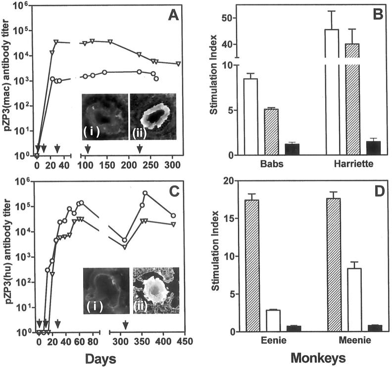
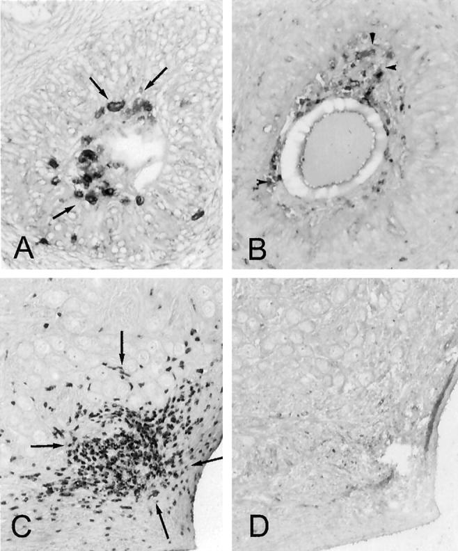
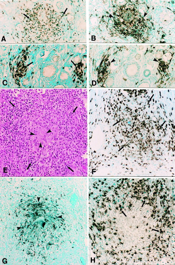
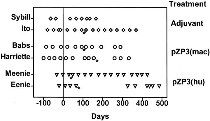
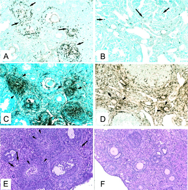
Autoimmune ovarian disease (AOD) is a probable cause of human premature ovarian failure, and a potential complication of contraceptive vaccines based on ovarian antigens. The diagnosis depends on detection of noninfectious ovarian inflammation (oophoritis) and serum antibody to ovarian and placental antigens. Mechanisms underlying AOD have been investigated in mice but not in primates. Herein, we report induction of AOD in primates, and compare the immunopathology between monkey and murine AOD. Four cynomolgus macaques immunized with monkey or human zona pellucida 3 peptide (pZP3) in adjuvant, developed T-cell responses to the immunizing peptide and produced antibody that bound to native zona pellucida in vivo. Immunostaining of ovaries from pZP3-immunized macaques showed numerous clusters of T cells co-localized with major histocompatibility complex II-positive macrophages in the ovarian interstitium. Such foci were not detected in untreated or adjuvant-treated control monkeys. This finding is comparable to murine pZP3-induced AOD. However, unlike murine AOD in which numerous granulomatous lesions are detected, severe granulomatous inflammation was detected in only one of three monkeys with abnormal immunohistology. Similar to mice with pZP3-induced AOD, the immunized monkeys retained normal ovarian function. The results are discussed in the context of complications of ZP-based human immunocontraceptive vaccines and case reports of human autoimmune oophoritis.


Figures

Figure 1.
Figure 1.
Immune response in female macaques immunized with pZP3(mac) (A and B) or pZP3(hu) (C and D). Serum antibody titers to the immunizing peptide were detected by enzyme-linked immunosorbent assay (A and C). Arrows indicate time of primary and booster immunizations. A: Antibody titers to pZP3(mac) [Babs (circles) and Harriette (triangles)]. Serum antibody from peptide-immunized Harriette recognized macaque zona pellucida (inset Aii) whereas sera from adjuvant-treated controls did not (inset Ai). In lymphocyte proliferation assays (B and D) animals immunized with pZP3(mac) (B) respond to the immunizing macaque peptide (open bars) and its human homologue (hatched bars) but not to the mouse peptide (solid bars). C: Antibody titers to pZP3(hu) [Meenie (circles) and Eenie (triangles)]. Direct immunofluorescence showed antibody bound to ZP (inset Cii) in Meenie but not in an adjuvant-treated macaque (inset Ci). In lymphocyte proliferative assays animals immunized with pZP3(hu) (D) respond to the immunizing peptide (hatched bars) and its homologous macaque peptide (open bars) but not to the dissimilar mouse peptide (solid bars). Data show stimulation index (±SEM) at 30 μmol/L concentration for each peptide. C is reprinted with permission from the Society of Reproduction, Inc. (Biol Reprod 1997, 56:767).
Figure 2.
Figure 2.
Immunoperoxidase staining for CD3+ T cells (arrows) and MHC II+ macrophages (arrowheads) in control macaque ovaries. A and B: Atretic follicles in adjacent sections have invading CD3+ T cells (A) along with MHC II-expressing cells (B). C and D: Adjacent sections of ovarian cortex reveal a cluster of CD3+ T cells (C) but absence of MHC II+ cells (D). Original magnifications, ×400.
Figure 3.
Figure 3.
Ovarian inflammation in pZP3(mac)- and pZP3(hu)-immunized macaques. A–D: Adjacent sections of a typical primate AOD lesion consisted of clusters of CD3+ T cells (A, arrows) and MHC II + cells (B, arrowheads). C and D: Adjacent sections of another typical primate AOD lesion stained for CD3 (C) and MHC II (D). Adjacent sections of a large granuloma in the ovary of Eenie that was immunized with pZP3(hu) in adjuvant. E: Typical granulomatous inflammation with eosinophilic center (arrowheads) surrounded by cuff of lymphocytes (arrows) (H&E). The granuloma contains many macrophage (Mac 387+) (F), a central core of MHC II-positive cells (G), and a rim of CD3+ T cells (H). Original magnifications, ×400.
Figure 4.
Figure 4.
Ovarian cyclicity in adjuvant-immunized (Sybill, Ito), pZP3(mac)-immunized (Harriette, Babs), and pZP3(hu)-immunized (Meenie, Eenie) macaques monitored until sacrifice. Each point indicates time of menstrual bleeding. Time of first immunization is indicated as day 0. Asterisk denotes surgical removal of one ovary.
Figure 5.
Figure 5.
Immunohistopathology of murine AOD. A, C, and E: Ovary of a mouse immunized with pZP3(mou) in CFA. Adjacent sections showing clusters of cells T cells (CD5+) (A, arrows) that co-localize with intensely MHC II+ cells (C, arrowheads). The opposite ovary stained with H&E (E) shows lymphocytic infiltration in the interstitial regions (arrows) and the ovarian follicles (arrowheads). B, D, and F: Ovary of an adjuvant-treated mouse showing a few T (CD5+) cells (B) and an adjacent section (D) with many MHC II+ macrophages in the interstitium and the corpus luteum. The opposite ovary (F) stained with H&E appears completely normal.

References

    1. Wheatcroft N, Weetman : Is premature ovarian failure an autoimmune disease? Autoimmunity 1997, 25:157–165 - PubMed
    1. Hoek A, Schoemaker J, Drexhage HA: Premature ovarian failure and ovarian autoimmunity. Endocr Rev 1997, 18:107-134 - PubMed
    1. Coulam CB, Kempers RD, Randall RV: Premature ovarian failure: evidence for the autoimmune mechanism. Fertil Steril 1981, 36:238-240 - PubMed
    1. Bannatyne P, Russel P, Shearman RP: Autoimmune oophoritis: a clinicopathological assessment of 12 cases. Int J Gynecol Pathol 1990, 9:191-207 - PubMed
    1. Russel P, Bannatyne P, Shearman RP: Premature hypergonadotropic ovarian failure: clinicopathologic study of 19 cases. Int J Gynecol Pathol 1982, 1:186-201 - PubMed

Publication types

MeSH terms