. 2002 Jan;160(1):195-204.
doi: 10.1016/s0002-9440(10)64363-5.

The alpha(1)beta(1) and alpha(2)beta(1) integrins provide critical support for vascular endothelial growth factor signaling, endothelial cell migration, and tumor angiogenesis

Affiliations

The alpha(1)beta(1) and alpha(2)beta(1) integrins provide critical support for vascular endothelial growth factor signaling, endothelial cell migration, and tumor angiogenesis

Donald R Senger et al. Am J Pathol. 2002 Jan.

Abstract

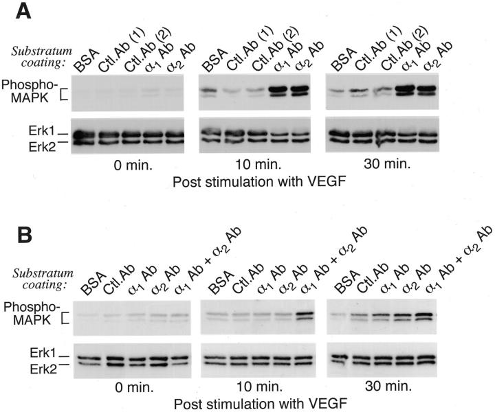
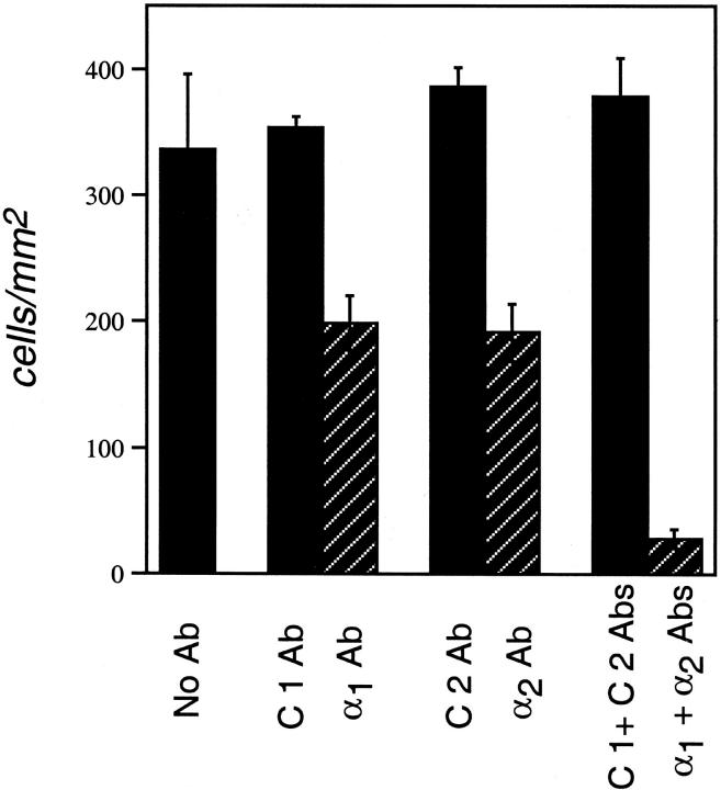
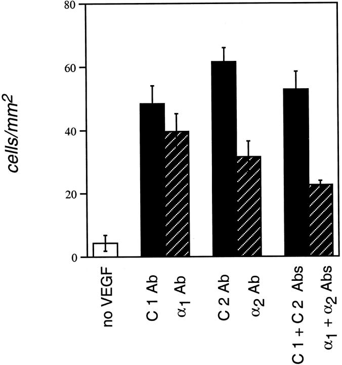
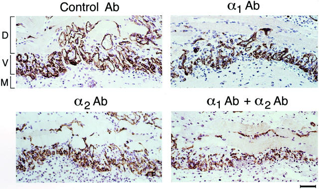
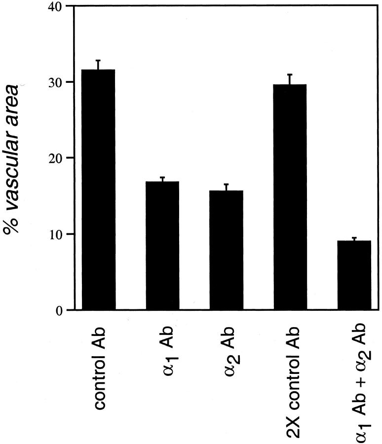
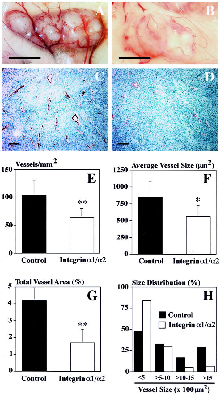
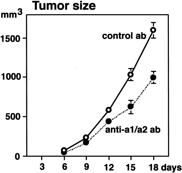
Angiogenesis is a complex process, involving functional cooperativity between cytokines and endothelial cell (EC) surface integrins. In this study, we investigated the mechanisms through which the alpha(1)beta(1) and alpha(2)beta(1) integrins support angiogenesis driven by vascular endothelial growth factor (VEGF). Dermal microvascular EC attachment through either alpha(1)beta(1) or alpha(2)beta(1) supported robust VEGF activation of the Erk1/Erk2 (p44/42) mitogen-activated protein kinase signal transduction pathway that drives EC proliferation. Haptotactic EC migration toward collagen I was dependent on alpha(1)beta(1) and alpha(2)beta(1) as was VEGF-stimulated chemotaxis of ECs in a uniform collagen matrix. Consistent with the functions of alpha(1)beta(1) and alpha(2)beta(1) in supporting signal transduction and EC migration, antibody antagonism of either integrin resulted in potent inhibition of VEGF-driven angiogenesis in mouse skin. Moreover, combined antagonism of alpha(1)beta(1) and alpha(2)beta(1) substantially reduced tumor growth and angiogenesis of human squamous cell carcinoma xenografts. Collectively, these studies identify critical collaborative functions for the alpha(1)beta(1) and alpha(2)beta(1) integrins in supporting VEGF signal transduction, EC migration, and tumor angiogenesis.


Figures

Figure 1.
Figure 1.
Adhesion of dermal microvascular ECs through either the α1β1 integrin or the α2β1 integrin supports VEGF activation of Erk1/Erk2 MAP kinases. Functional integrin Abs or control Abs were immobilized on plastic and remaining protein-binding sites were blocked with BSA, as described in Materials and Methods. After plating, cells attached and spread on substratum coated with α1 Ab or α2 Ab, but remained unattached to wells coated with isotype control Abs or BSA alone. After plating, MAPK phosphorylation was allowed to decay for 3 hours before stimulation with VEGF. A: Wells coated with 10 μg/ml Ab. As indicated by staining with phospho-specific MAPK Abs, VEGF induced marked activation in cells plated on α1 Ab or α2 Ab but poorly induced MAPK activation in cells plated on control Abs or BSA. Staining with Ab that recognizes both phosphorylated and nonphosphorylated forms of Erk1 and Erk2 established that they were present equally in all samples. B: VEGF more efficiently induced activation of MAPK in cells plated on substratum coated with a combination of α1 Ab and α2 Ab (0.2 μg/ml of each) than in cells plated on substratum coated with each Ab alone (0.2 μg/ml). For all experiments (A and B), failure of VEGF to activate MAPK in cells plated on BSA or control (Ctl) Abs was not attributable to anoikis because cell viability remained >90%, as determined by replating of cells on collagen-coated plastic.
Figure 2.
Figure 2.
The α1β1 and α2β1 integrins each support dermal microvascular EC-directed migration toward collagen I (haptotaxis). Cells were incubated with integrin-blocking or control Abs, and then placed in Transwell migration chambers containing filters coated on the undersides with collagen I. α1 Ab and α2 Ab were each inhibitory, but isotype control Abs (C1 Ab, C2 Ab) were without effect, indicating that both α1β1 and α2β1 function in migration toward collagen I. Error bars indicate standard deviations.
Figure 3.
Figure 3.
Antagonism of α1β1 and α2β1 integrins suppresses dermal microvascular EC chemotaxis toward VEGF. Cells were incubated with integrin-blocking or matched isotype control Abs and then placed in Transwell migration chambers containing filters coated uniformly on both sides with collagen I. To stimulate chemotaxis, VEGF was added to the lower chamber; note that migration was relatively insignificant in the absence of VEGF (left, single open column). Error bars indicate standard deviations.
Figure 4.
Figure 4.
Inhibition of VEGF-driven angiogenesis in mouse skin by α1 Ab and α2 Ab, as visualized by CD31 Ab staining of sections cut from paraffin-embedded specimens. New blood vessels (V) at the interface between the Matrigel implant containing the angiogenic stimulus (M) and the overlying dermis and smooth muscle cell layer (D) are stained for CD31 (brown color). Note reduced blood vessel diameters and reduced percentage of vascular cross-sectional area in integrin Ab groups in comparison with control. Scale bar, 50 μm.
Figure 5.
Figure 5.
Quantitation of angiogenesis inhibition by α1 Ab and α2 Ab in mouse skin. Vascular cross-sectional area as a percentage of total tissue area was measured at the interface between dermis and the angiogenic stimulus (see Figure 4 ▶ , above) as described in Materials and Methods. Data are presented as the mean ± SEM. Total cross-sectional area of new blood vessels in the α1 Ab and α2 Ab treatment groups were each reduced ∼45% relative to controls (P < 0.001). Administration of α1 Ab together with α2 Ab resulted in further inhibition of neovascularization, yielding an ∼70% reduction (P < 0.001).
Figure 6.
Figure 6.
Combined treatment with α1 Ab plus α2 Ab inhibits tumor angiogenesis. Rarefaction of blood vessels in human A431 tumors from nude mice treated with α1 Ab plus α2 Ab (B), as compared with control Ab (A) (scale bar, 5 mm). Immunostaining with an anti-CD31 mAb demonstrated rarefaction and decreased size of tumor blood vessels in the α1 Ab plus α2 Ab treatment group (D) as compared to controls (C) (scale bar, 100 μm). E–H: Quantitative, computer-assisted image analysis revealed a significant inhibition of angiogenesis in A431 tumors from animals treated with α1 Ab plus α2 Ab (P < 0.01), as measured by the number of blood vessels per mm tumor cross-sectional area (E). Furthermore, in A431 tumors from animals treated with α1 Ab plus α2 Ab, there was a significant reduction (P < 0.05) in average vessel size (F) with strong reduction in number of vessels with cross-sectional area >1000 μm (H). G: Overall, blood vessel area in cross-section as a percentage of total tumor area was reduced by ∼60% (P < 0.01) in A431 tumors from animals treated with α1 Ab plus α2 Ab, as compared with control Ab. CD31-stained blood vessels were evaluated in three different ×10 fields in sections obtained from five different tumors for each group.
Figure 7.
Figure 7.
Combined treatment with α1 Ab plus α2 Ab inhibits tumor growth. Administration of α1 Ab plus α2 Ab (closed circles) significantly (P < 0.01) inhibited intradermal tumor growth of human A431 cells, as compared with control Ab (open circles). Values represent mean values ± SEM for 10 tumors for each treatment group and time point. Note that the Abs used recognize mouse but not human integrins, and therefore Ab did not bind A431 tumor cells. Thus, effects of integrin Abs were limited to the mouse host.

Similar articles

Cited by

References

    1. Senger DR, Van de Water L, Brown LF, Nagy JA, Yeo KT, Yeo TK, Berse B, Jackman RW, Dvorak AM, Dvorak HF: Vascular permeability factor (VPF, VEGF) in tumor biology. Cancer Metastasis Rev 1993, 12:303-324 - PubMed
    1. Ferrara N: The role of vascular endothelial growth factor in pathological angiogenesis. Breast Cancer Res Treat 1995, 36:127-137 - PubMed
    1. Dvorak HF, Brown LF, Detmar M, Dvorak AM: Vascular permeability factor/vascular endothelial growth factor, microvascular hyperpermeability, and angiogenesis. Am J Pathol 1995, 146:1029-1039 - PMC - PubMed
    1. Mustonen T, Alitalo K: Endothelial receptor tyrosine kinases involved in angiogenesis. J Cell Biol 1995, 129:895-898 - PMC - PubMed
    1. Ferrara N, Henzel WJ: Pituitary follicular cells secrete a novel heparin-binding growth factor specific for vascular endothelial cells. Biochem Biophys Res Commun 1989, 161:851-858 - PubMed

Publication types

MeSH terms