. 2002 Jan;135(1):257-65.
doi: 10.1038/sj.bjp.0704439.

Gabapentin inhibits high-threshold calcium channel currents in cultured rat dorsal root ganglion neurones

Affiliations

Gabapentin inhibits high-threshold calcium channel currents in cultured rat dorsal root ganglion neurones

K G Sutton et al. Br J Pharmacol. 2002 Jan.

Abstract

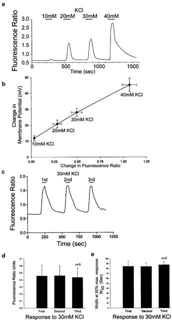
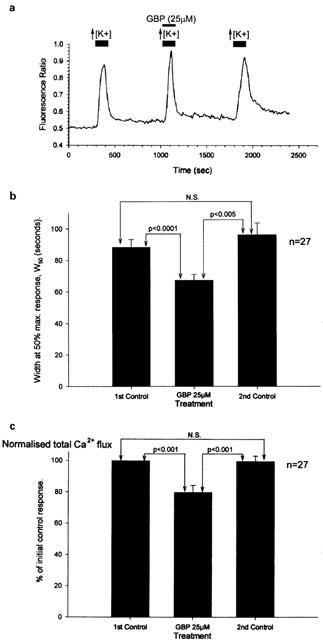
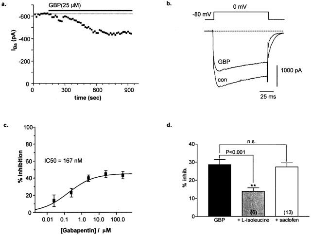
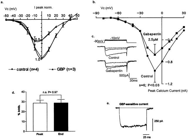
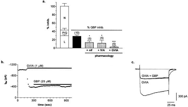
1. This study examined the action of gabapentin (gabapentin,1-(aminomethyl) cyclohexane acetic acid (Neurontin) on voltage-gated calcium (Ca(2+)) channel influx recorded in cultured rat dorsal root ganglion (DRG) neurones. 2. Voltage-gated Ca(2+) influx was monitored using both fura-2 based fluorescence Ca(2+) imaging and the whole-cell patch clamp technique. 3. Imaging of intracellular Ca(2+) transients revealed that gabapentin inhibited KCl (30 mM)-evoked voltage-dependent Ca(2+) influx. Both the duration for 50% of the maximum response (W50) and total Ca(2+) influx were significantly reduced by approximately 25-30% in the presence of gabapentin (25 microM). 4. Gabapentin potently inhibited the peak whole-cell Ca(2+) channel current (I(Ba)) in a dose-dependent manner with an estimated IC(50) value of 167 nM. Block was incomplete and saturated at a maximal concentration of 25 microM. 5. Inhibition was significantly decreased in the presence of the neutral amino acid L-isoleucine (25 microM) but unaffected by application of the GABA(B) antagonist, saclofen (200 microM), suggesting a direct action on the alpha(2)delta subunit of the Ca(2+) channel. 6. Gabapentin inhibition was voltage-dependent, producing an approximately 7 mV hyperpolarizing shift in current voltage properties and reducing a non-inactivating component of whole-cell current activated at relatively depolarized potentials. 7. The use of specific Ca(2+) channel antagonists revealed a mixed pharmacology of the gabapentin-sensitive current (N-, L- and P/Q-type), which is dominated by N-type current. 8. The present study is the first to demonstrate that gabapentin directly mediates inhibition of voltage-gated Ca(2+) influx in DRG neurones, providing a potential means for gabapentin to effectively mediate spinal anti-nociception.


Figures

Figure 1
Figure 1
(a) Trace showing changes in fluorescence ratio produced by changes in KCl in imaging experiments. (b) Graph showing linear correlation between change in fluorescence ratio and membrane potential at varying concentrations of K+. The plot was produced from mean data±s.e.mean obtained from imaging experiments (n=19) and measuring membrane potential changes (n=6 – 7); R2=0.9977. (c) Trace from an individual DRG neurone showing that, three pulses of 30 mM KCl gave repeatable responses that fully recovered between stimulation. (d and e) Bar graphs show that consistent control Ca2+ transients were obtained in response to repeated stimuli of 30 mM KCl applied to the same cells. Data plotted are the mean response as a fluorescence ratio or duration at 50% of the transient amplitude.
Figure 2
Figure 2
Gabapentin inhibited Ca2+ influx into cultured DRG as measured using fura-2 fluorescence imaging. (a) Trace from a single cultured DRG neurone showing a decrease in response duration and total Ca2+ flux by gabapentin (25 μM), but no significant change in the amplitude of the peak Ca2+ transient. (b and c) Bar charts showing the inhibitory actions of 25 μM gabapentin (GBP) on the duration of the Ca2+ transients evoked by 30 mM KCl measured at 50% of the peak amplitude (W50) and the total Ca2+ flux normalized with respect to the first control Ca2+ transient.
Figure 3
Figure 3
(a) Time-course showing gabapentin inhibition of peak IBa. (b) Whole-cell current trace of peak HVA IBa activated under control conditions (con) and following a 10 min application of gabapentin (GBP). (c) Dose-response of GBP-mediated inhibition of peak HVA IBa. Data points represent mean±s.e.mean (n=3 – 8 determinants) and were fitted with the following sigmoidal equation: Y=max/1+(IC50/[GBP]n) where n is the Hill slope factor=0.55, max is maximum inhibition=46.8% and IC50=167 nM. (d) Bar graph (mean±s.e.mean) showing that inhibition of peak HVA IBa by gabapentin (GBP) is significantly reduced in the presence of L-isoleucine (25 μM) but is unaffected following treatment with saclofen (200 μM, n=10, 6 and 13, respectively).
Figure 4
Figure 4
(a) Mean±s.e.mean ramp-activated whole-cell currents (0.9 ms/mV) from −50 mV under control conditions (n=4) and following a 10 min application of gabapentin (25 μM; n=3). Solid line indicates Boltzmann fit to the data. (b) Mean (±s.e.mean; n=6 determinants) I/V relationship for whole-cell voltage-gated Ca2+ currents (2 mM Ca2+) recorded under control conditions and following a 3 – 5 min application of gabapentin by low pressure ejection (2.5 μM; P<0.03). (c) inset records showing Ca2+ currents recorded in the absence or presence of gabapentin (Vc=−30, −20 and 10 mV). (d) Bar graph of mean inhibition (±s.e.mean; n=10 determinants) of HVA IBa measured at the peak vs the end of the current step. (e) Gabapentin-sensitive difference current obtained by subtracting peak HVA current traces recorded under control conditions and following a 10 min application of gabapentin (25 μM).
Figure 5
Figure 5
(a) Pharmacology of gabapentin-sensitive peak HVA IBa (mean±s.e.mean). Clear bars represent mean data for inhibition of peak HVA IBa with Ca2+ channel antagonists (L-, P/Q- or N-type; n=6, 8 and 7, respectively). The black bar represents mean total current inhibited by gabapentin. Grey bars represent mean gabapentin-sensitive current remaining in the presence of L- (+nif), P/Q- (+IVA) or N- (+GVIA) type blockers. (b) Time-course showing gabapentin inhibition of peak IBa is virtually eliminated following block of the N-type current with ω-CgTx GVIA. (c) Whole-cell current trace of peak HVA IBa activated under control conditions and following sequential applications of ω-CgTx GVIA (1 μM, GVIA) followed by gabapentin (25 μM) in the presence of GVIA (GVIA+GBP).

References

    1. BACKONJA M., BEYDOUN A., EDWARDS K.R., SCHWARTZ S.L., FONESCA V., HES M., LAMOREAUX L., GAROFALO E. Gabapentin for the symptomatic treatment of painful neuropathy in patients with diabetes mellitus. JAMA. 1998;280:1831–1836. - PubMed
    1. BROWN J.P., GEE N.S. Cloning and deletion mutagenesis of the α2δ calcium channel subunit from porcine cerebral cortex. J. Biol. Chem. 1998;273:25458–25465. - PubMed
    1. CARACENI A., ZECCA E., MARTINI C., DE CONNO F. Gabapentin as an adjuvant to opioid analgesia for neuropathic cancer pain. J. Pain. Symp. Manag. 1999;17:441–445. - PubMed
    1. CESENA R.M., CALCUTT N.A. Gabapentin prevents hyperalgesia during the formalin test in diabetic rats. Neurosci. Lett. 1999;262:101–104. - PubMed
    1. CHENG J-K,. , PAN H-L., EISENACH J.C. Antiallodynic effect of intrathecal gabapentin and its interaction with clonidine in a rat model of postoperative pain. Anesthesiology. 2000;92:1126–1131. - PubMed

Publication types

MeSH terms