. 2002 Jan 15;30(2):532-44.
doi: 10.1093/nar/30.2.532.

Convergence and constraint in eukaryotic release factor 1 (eRF1) domain 1: the evolution of stop codon specificity

Affiliations

Convergence and constraint in eukaryotic release factor 1 (eRF1) domain 1: the evolution of stop codon specificity

Yuji Inagaki et al. Nucleic Acids Res. .

Abstract

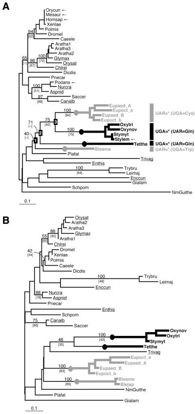
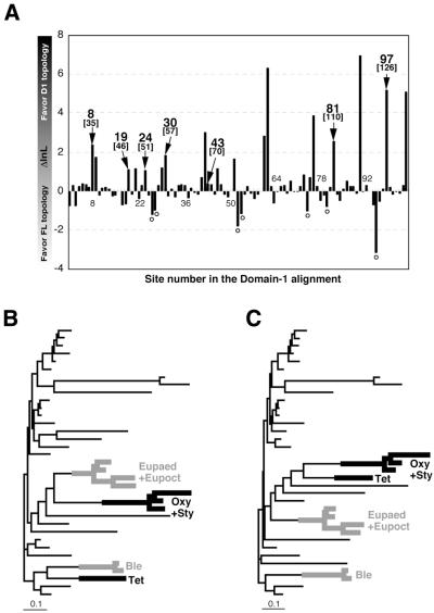
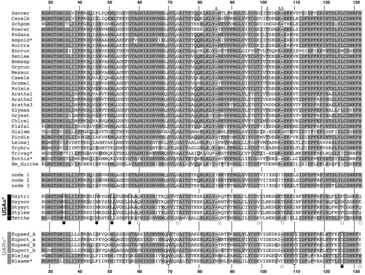
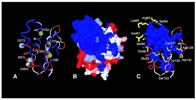
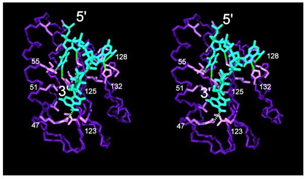
Class 1 release factor in eukaryotes (eRF1) recognizes stop codons and promotes peptide release from the ribosome. The 'molecular mimicry' hypothesis suggests that domain 1 of eRF1 is analogous to the tRNA anticodon stem-loop. Recent studies strongly support this hypothesis and several models for specific interactions between stop codons and residues in domain 1 have been proposed. In this study we have sequenced and identified novel eRF1 sequences across a wide diversity of eukaryotes and re-evaluated the codon-binding site by bioinformatic analyses of a large eRF1 dataset. Analyses of the eRF1 structure combined with estimates of evolutionary rates at amino acid sites allow us to define the residues that are under structural (i.e. those involved in intramolecular interactions) versus non-structural selective constraints. Furthermore, we have re-assessed convergent substitutions in the ciliate variant code eRF1s using maximum likelihood-based phylogenetic approaches. Our results favor the model proposed by Bertram et al. that stop codons bind to three 'cavities' on the protein surface, although we suggest that the stop codon may bind in the opposite orientation to the original model. We assess the feasibility of this alternative binding orientation with a triplet stop codon and the eRF1 domain 1 structures using molecular modeling techniques.


Figures

Figure 1
Figure 1
(A) An unrooted eRF1 tree based on the FL dataset using the Γ-FITCH protein distance method. Abbreviations of the sequence names are listed in Table 2. BP are listed for major nodes. Percent occurrence in 10 000 quartet puzzling trees are given in brackets. Dashes indicate that given nodes were not supported by bootstrap or quartet puzzling analyses. The novel sequences determined or identified in this study are underlined. The redundant sequences removed from the analyses using the D1 dataset are marked by arrows. The ciliate lineages that independently converted the standard code to variant codes [UGA = * (UAR = Gln) and UAR = * (UGA = Cys or Trp)] are highlighted by thick black and gray lines, respectively. (B) An unrooted eRF1 tree based on the D1 dataset using the Γ-FITCH protein distance method.
Figure 2
Figure 2
(A) Comparison of site 1nL calculated under the FL and D1 tree topologies. lnL scores calculated from the D1 dataset under the D1 and FL topologies (see Fig. 1A and B) were compared on a site-by-site basis. The sites that were inferred to have evolved convergently in the eRF1s from Oxytricha + Stylonychia and Tetrahymena (UGA = *) and Euplotes and Blepharisma (UAR = *) are highlighted by arrows with site numbers in the alignment (the human eRF1 numbering is given in parentheses). A Γ-FITCH tree based on the D1 dataset excluding the seven convergent sites (B) and a tree based on the D1 dataset excluding the seven sites with negative ΔlnL scores (marked with open circles in Fig. 2A) (C). Details of the analyses are described in the text. Sequence names except for ciliates are omitted. Oxy, Oxytricha; Sty, Stylonychia; Tet, Tetrahymena; Eupaed, Euplotes aediculatus; Eupoct, Euplotes octocarinatus; Ble, Blepharisma. The ciliate lineages that independently converted the standard code to variant codes [UGA = * (UAR = Gln) and UAR = * (UGA = Cys or Trp)] are highlighted by thick black and gray lines, respectively.
Figure 3
Figure 3
An amino acid alignment of eRF1 D1. Abbreviations of the sequence names are listed in Table 2. Residues conserved among >70% of standard code eRF1s are shaded. Converged unvaried sites in ciliate eRF1s that are identified both in this study and the study by Lozupone et al. (23) are boxed with filled circles. Converged unvaried sites newly identified in this study are boxed. ‘Convergent’ sites identified only in the study of Lozupone et al. (23) are marked with open circles. The sequences labeled as nodes 1–3 are the ancestral ciliate sequences inferred for these nodes on the full-length topology (see Fig. 1A). The residue numbers are based on human eRF1 (GenBank accession no. U90176). Asterisks indicate partial sequences. Sites that were excluded from the computational analyses are indicated by open triangles. The amino acid residues in the ancestral ciliate eRF1s (nodes 1–3 in Fig. 1A) for these excluded sites are not available and are indicated by question marks.
Figure 4
Figure 4
Distribution of site rates in eRF1 D1 and converged unvaried sites in the UGA = * and UAR = * eRF1s mapped on the human eRF1 D1 structure (PDB accession no. 1DT9). The five site rates categories are color coded from blue (slowest) to red (fastest). (A) Distribution of the converged unvaried sites mapped on the human eRF1 D1 structure. Gray spheres indicate a convergence in the ciliate VC-eRF1s with the UAR = * code (Euplotes and Blepharisma) and green spheres a convergence in the VC-eRF1s with the UGA = * code (Oxytricha, Stylonychia and Tetrahymena). The labels indicate the identity of the ancestral residue on the left of the number and the convergent change on the right of the number. Numbering is based on human eRF1 (GenBank accession no. U90176). (B) Site rates of D1 of eRF1 mapped on the solvent-accessible surface. The surface cavities are labeled as per Bertram et al. (10). (C) Distribution of the residues included in the slowest category. All side chains potentially performing a structural function (see criteria in the text) are represented as a molecular surface. Polar residues for which no structural function could be assigned are displayed in yellow.
Figure 5
Figure 5
Stereoview of stop codon 5′-UAA-3′ (blue) bound to the human release factor eRF1 N-terminal domain (purple) in the reverse orientation to the previously proposed model (10). Only the backbone atoms of eRF1 and the side chain interacting with the trinucleotide are displayed. Green dotted lines indicate possible dipolar interactions conferring binding specificity to the stop codon.

References

    1. Nakamura Y. and Ito,K. (1998) How proteins read the stop codon and terminate translation. Genes Cells, 3, 265–278. - PubMed
    1. Kisselev L.L. and Buckingham,R.H. (2000) Translational termination comes of age. Trends Biochem. Sci., 25, 561–566. - PubMed
    1. Poole E. and Tate,W. (2000) Release factors and their role as decoding proteins: specificity and fidelity for termination of protein synthesis. Biochim. Biophys. Acta, 1493, 1–11. - PubMed
    1. Bertram G., Innes,S., Minella,O., Richardson,J. and Stansfield,I. (2001) Endless possibilities: translation termination and stop codon recognition. Microbiology, 147, 255–269. - PubMed
    1. Dontsova M., Frolova,L., Vassilieva,J., Piendl,W., Kisselev,L. and Garber,M. (2000) Translation termination factor aRF1 from the archaeon Methanococcus jannaschii is active with eukaryotic ribosomes. FEBS Lett., 472, 213–216. - PubMed

Publication types

MeSH terms

Associated data