

An evaluation of tyramide signal amplification and archived fixed and frozen tissue in microarray gene expression analysis
- PMID: 11788730
- PMCID: PMC99843
- DOI: 10.1093/nar/30.2.e4
An evaluation of tyramide signal amplification and archived fixed and frozen tissue in microarray gene expression analysis
Abstract
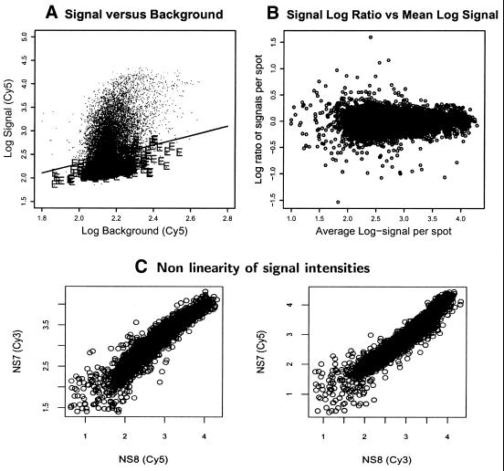
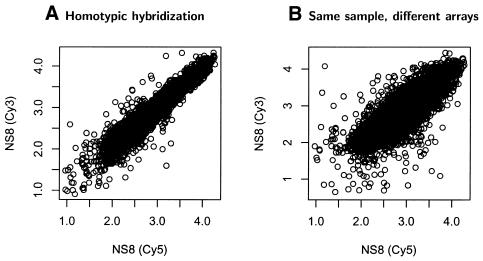
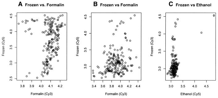
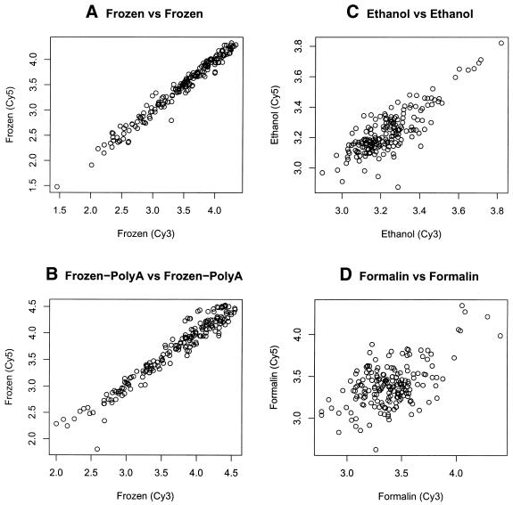
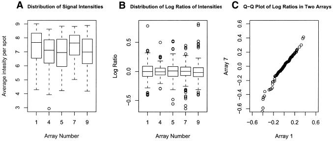
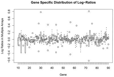
Archival formalin-fixed, paraffin-embedded and ethanol-fixed tissues represent a potentially invaluable resource for gene expression analysis, as they are the most widely available material for studies of human disease. Little data are available evaluating whether RNA obtained from fixed (archival) tissues could produce reliable and reproducible microarray expression data. Here we compare the use of RNA isolated from human archival tissues fixed in ethanol and formalin to frozen tissue in cDNA microarray experiments. Since an additional factor that can limit the utility of archival tissue is the often small quantities available, we also evaluate the use of the tyramide signal amplification method (TSA), which allows the use of small amounts of RNA. Detailed analysis indicates that TSA provides a consistent and reproducible signal amplification method for cDNA microarray analysis, across both arrays and the genes tested. Analysis of this method also highlights the importance of performing non-linear channel normalization and dye switching. Furthermore, archived, fixed specimens can perform well, but not surprisingly, produce more variable results than frozen tissues. Consistent results are more easily obtainable using ethanol-fixed tissues, whereas formalin-fixed tissue does not typically provide a useful substrate for cDNA synthesis and labeling.
Figures
Similar articles
-
Preparation of archival formalin-fixed paraffin-embedded mouse liver samples for use with the Agilent gene expression microarray platform.J Pharmacol Toxicol Methods. 2013 Sep-Oct;68(2):260-268. doi: 10.1016/j.vascn.2013.02.008. Epub 2013 Mar 1. J Pharmacol Toxicol Methods. 2013. PMID: 23458726
-
Methods comparison for high-resolution transcriptional analysis of archival material on Affymetrix Plus 2.0 and Exon 1.0 microarrays.Biotechniques. 2009 Jul;47(1):587-96. doi: 10.2144/000113169. Biotechniques. 2009. PMID: 19594443
-
Illumina whole-genome complementary DNA-mediated annealing, selection, extension and ligation platform: assessing its performance in formalin-fixed, paraffin-embedded samples and identifying invasion pattern-related genes in oral squamous cell carcinoma.Hum Pathol. 2011 Dec;42(12):1911-22. doi: 10.1016/j.humpath.2011.02.011. Epub 2011 Jun 17. Hum Pathol. 2011. PMID: 21683979
-
Tyramide signal amplification for DNA and mRNA in situ hybridization.Methods Mol Biol. 2006;326:33-60. doi: 10.1385/1-59745-007-3:33. Methods Mol Biol. 2006. PMID: 16780193 Review.
-
Tissue handling and specimen preparation in surgical pathology: issues concerning the recovery of nucleic acids from formalin-fixed, paraffin-embedded tissue.Arch Pathol Lab Med. 2008 Dec;132(12):1929-35. doi: 10.5858/132.12.1929. Arch Pathol Lab Med. 2008. PMID: 19061293 Review.
Cited by
-
Whole-genome gene expression profiling of formalin-fixed, paraffin-embedded tissue samples.PLoS One. 2009 Dec 3;4(12):e8162. doi: 10.1371/journal.pone.0008162. PLoS One. 2009. PMID: 19997620 Free PMC article.
-
A new paradigm for biospecimen banking in the personalized medicine era.Am J Clin Pathol. 2011 Nov;136(5):679-84. doi: 10.1309/AJCP7DWCQ1SWJTWU. Am J Clin Pathol. 2011. PMID: 22031304 Free PMC article.
-
Identification of molecular mechanisms for cellular drug resistance by combining drug activity and gene expression profiles.Br J Cancer. 2005 Aug 22;93(4):483-92. doi: 10.1038/sj.bjc.6602699. Br J Cancer. 2005. PMID: 16012520 Free PMC article.
-
Microarray analysis of RNA extracted from formalin-fixed, paraffin-embedded and matched fresh-frozen ovarian adenocarcinomas.BMC Med Genomics. 2009 May 8;2:23. doi: 10.1186/1755-8794-2-23. BMC Med Genomics. 2009. PMID: 19426511 Free PMC article.
-
Global transcriptome analysis of genetically identified neurons in the adult cortex.J Neurosci. 2006 Sep 27;26(39):9956-66. doi: 10.1523/JNEUROSCI.0468-06.2006. J Neurosci. 2006. PMID: 17005859 Free PMC article.
References
-
- Lockhart D.J., Dong,H., Byrne,M.C., Follettie,M.T., Gallo,M.V., Chee,M.S., Mittmann,M., Wang,C., Kobayashi,M., Horton,H. et al. (1996) Expression monitoring by hybridization to high-density oligonucleotide arrays. Nat. Biotechnol., 14, 1675–1680. - PubMed
-
- DeRisi J.L., Iyer,V.R. and Brown,P.O. (1997) Exploring the metabolic and genetic control of gene expression on a genomic scale. Science, 278, 680–686. - PubMed
-
- Speel E.J., Hopman,A.H. and Komminoth,P. (1999) Amplification methods to increase the sensitivity of in situ hybridization: play card(s). J. Histochem. Cytochem., 47, 281–288. - PubMed
Publication types
MeSH terms
Substances
Grants and funding
LinkOut - more resources
Full Text Sources
Other Literature Sources
Research Materials
Miscellaneous
