Comparative Study
. 2002 Feb;184(3):654-65.
doi: 10.1128/JB.184.3.654-665.2002.

Characterization of indigoidine biosynthetic genes in Erwinia chrysanthemi and role of this blue pigment in pathogenicity

Affiliations
Comparative Study

Characterization of indigoidine biosynthetic genes in Erwinia chrysanthemi and role of this blue pigment in pathogenicity

Sylvie Reverchon et al. J Bacteriol. 2002 Feb.

Abstract

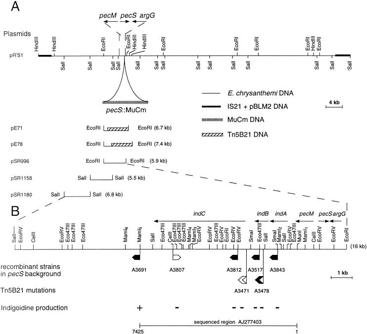
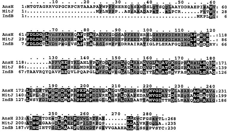
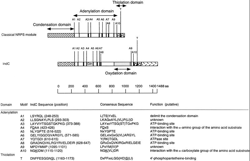
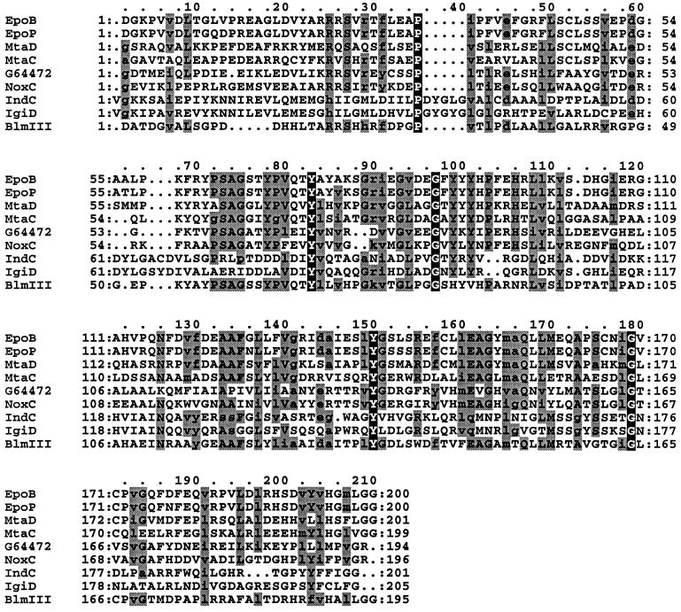
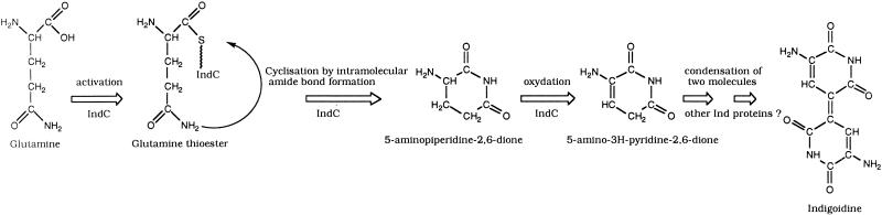
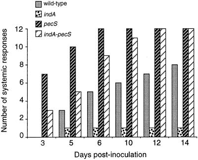
In the plant-pathogenic bacterium Erwinia chrysanthemi production of pectate lyases, the main virulence determinant, is modulated by a complex network involving several regulatory proteins. One of these regulators, PecS, also controls the synthesis of a blue pigment identified as indigoidine. Since production of this pigment is cryptic in the wild-type strain, E. chrysanthemi ind mutants deficient in indigoidine synthesis were isolated by screening a library of Tn5-B21 insertions in a pecS mutant. These ind mutations were localized close to the regulatory pecS-pecM locus, immediately downstream of pecM. Sequence analysis of this DNA region revealed three open reading frames, indA, indB, and indC, involved in indigoidine biosynthesis. No specific function could be assigned to IndA. In contrast, IndB displays similarity to various phosphatases involved in antibiotic synthesis and IndC reveals significant homology with many nonribosomal peptide synthetases (NRPS). The IndC product contains an adenylation domain showing the signature sequence DAWCFGLI for glutamine recognition and an oxidation domain similar to that found in various thiazole-forming NRPS. These data suggest that glutamine is the precursor of indigoidine. We assume that indigoidine results from the condensation of two glutamine molecules that have been previously cyclized by intramolecular amide bond formation and then dehydrogenated. Expression of ind genes is strongly derepressed in the pecS background, indicating that PecS is the main regulator of this secondary metabolite synthesis. DNA band shift assays support a model whereby the PecS protein represses indA and indC expression by binding to indA and indC promoter regions. The regulatory link, via pecS, between indigoidine and virulence factor production led us to explore a potential role of indigoidine in E. chrysanthemi pathogenicity. Mutants impaired in indigoidine production were unable to cause systemic invasion of potted Saintpaulia ionantha. Moreover, indigoidine production conferred an increased resistance to oxidative stress, indicating that indigoidine may protect the bacteria against the reactive oxygen species generated during the plant defense response.


Figures

FIG. 1.
FIG. 1.
(A) Restriction map of plasmid pR’S1 harboring the argG, pecS::MudIIPR13, pecM, and indABC region of E. chrysanthemi 3937. (B) Insertion mutagenesis of the indABC region of E. chrysanthemi 3937. The sites of insertion of uidA-Km cassettes are indicated by small flags. Localization of the Tn5-B21 insertions is indicated by larger flags. In the sequenced region AJ277403, the exact positions of Tn5-B21 insertions are at nucleotide 1619 for A3478 and nucleotide 2286 for A3471. These positions were deduced from sequences obtained from plasmids pE71 and pE78 using primer Tn5 (5′GGGAAAGGTTCCGTTCAGGACG3′), which hybridizes at the end of transposon Tn5-B21. The transcription direction of the reporter gene is shown by the orientation of the flags. Where the reporter (uidA or lacZ) gene is expressed, the flag is black. The phenotypes of the mutants obtained after recombination of these insertions into the E. chrysanthemi pecS chromosome is described by the + or − signs. The deduced transcriptional organization of this DNA region is indicated by arrows.
FIG. 2.
FIG. 2.
Alignment of E. chrysanthemi IndB protein with phosphatases involved in antibiotic synthesis. The proteins represented are AnsH from S. collinus (AF131879) and MitJ from S. lavendulae (AF127374). The residues conserved in all the three proteins are in black boxes. The residues conserved in two of the three proteins are in grey boxes. Conservative substitutions are in lowercase letters. Dots denote gaps.
FIG. 3.
FIG. 3.
Structural organization of the IndC peptide synthetase. The relative locations of the highly conserved signature sequences found in adenylation (A1 to A10) and thiolation (T) domains are marked as stripes. Their amino acid sequences, in one-letter code, and their putative functions are indicated. Alternative amino acids for a particular position are shown in parentheses (x, any amino acid). The residues D and K underlined in motifs A4 and A10, respectively, mediate electrostatic interactions with the α-amino and α-carboxylate group of the amino acid substrate. The residue S underlined in motif T serves as the covalent attachment point for the 4′-phosphopantetheine cofactor of NRPS. This attachment is catalyzed by enzymes belonging to the superfamily of 4′-phosphopantetheine transferases. These enzymes promote the nucleophilic attack of the invariant serine hydroxy group to the pyrophosphate bridge of coenzyme A, resulting in a transfer of the 4′-phosphopantetheine cofactor to the T domain and a liberation of 3′,5′-ADP.
FIG. 4.
FIG. 4.
Amino acid comparison of putative oxidation domains. The amino acid positions of each putative oxidation domain are 1023 to 1222 for EpoB and EpoP from S. cellulosum, 1042 to 1242 for MtaD and 1083 to 1281 for MtaC from S. aurantiaca, 861 to 1061 for IndC from E. chrysanthemi, 665 to 869 for IgiD from V. indigofera, 734 to 928 for BlmIII from S. verticillus, 37 to 232 for NoxC from P. abyssi, and 2 to 195 for the hypothetical protein G64472 from M. jannaschii. The residues conserved in all proteins are in black boxes. The residues conserved in 60% of the proteins are in grey boxes. Conservative substitutions are in lowercase letters. Dots denote gaps.
FIG. 5.
FIG. 5.
Proposed model for indigoidine biosynthesis. IndC is responsible for glutamine activation as its thioester. Glutamine is then cyclized by the formation of an intramolecular amide bond. The resulting molecule, 5-aminopiperidine-2,6-dione, is then dehydrogenated, probably by the oxidation domain of IndC, to generate 5-amino-3H-pyridine-2,6-dione. Condensation of two such molecules by other Ind proteins could then generate indigoidine.
FIG. 6.
FIG. 6.
Expression of indA::uidA and indC::uidA fusions in Saintpaulia plants and in M63 glycerol synthetic medium. For in planta expression, β-glucuronidase activities were determined in bacterial cells collected from inoculated plant leaves when the first symptoms occurred. The data represent one of two separate experiments which gave similar results.
FIG. 7.
FIG. 7.
(A) Identification of the E. chrysanthemi indA and indC transcription start site. RNAs (10 μg [lane 1] or 20 μg [lane 2]) from E. chrysanthemi A1524 cells grown on LB medium were submitted to primer extension analysis using indA- and indC-specific primers. DNA sequencing ladders were generated with the same primers (lanes A, C, G, and T). The nucleotide sequences of both the coding and noncoding strands are shown on the right. Arrows indicate the position of the specific transcription initiation sites (on the left). (B) Nucleotide sequences of the indA and indC regulatory regions. Sequences are numbered from the transcription start sites. The stop codon of the preceding gene, initiation codon, the ribosome-binding sites (S.D.), and regions corresponding to the −10 and −35 promoter sites are underlined. The transcriptional start sites are double underlined. (C) Interaction of the purified PecS protein with the E. chrysanthemi indA and indC regulatory regions. End-labeled and purified DNA fragments containing the indA and indC regulatory regions were incubated with various concentrations of the PecS protein.
FIG. 8.
FIG. 8.
Hydrogen peroxide sensitivities of the wild-type E. chrysanthemi 3937, pecS mutant A3953, indA mutant A3954, and the double indA-pecS mutant A3956. (A) Experiments were performed by incubating 3 × 10⁹ bacteria with 10 mM hydrogen peroxide for the times indicated and determining viable cells by serial dilution and plating on LB agar. Survival of the H2O2-treated cells was normalized to the number of CFU at the beginning of the challenge. (B) The same experiments were performed with bacteria preincubated with either H2O2 (500 μM) or paraquat (40 μM). The data presented were obtained with H2O2 preinduction, but similar results were obtained with paraquat preinduction (data not shown). The data represent one of three separate experiments which gave similar results.
FIG. 9.
FIG. 9.
Development of symptoms caused by E. chrysanthemi 3937 (wild type) and its indA, pecS, and indA-pecS derivatives on S. ionantha. The y axis indicates the number of plants (of 12 tested) with at least complete maceration of the inoculated leaf with its petiole. One representative experiment as described in Materials and Methods is shown. Other details are indicated in the text.

Similar articles

Cited by

References

    1. Altschul, A. F., W. Gish, W. Miller, E. W. Myers, and D. J. Lipman. 1990. Basic local alignment search tool. J. Mol. Biol. 215:403–410. - PubMed
    1. Ausubel, F. M., R. Brent, R. E. Kingston, D. D. Moore, J. G. Seidman, J. A. Smith, and K. Struhl (ed.). 1987. Current protocols in molecular biology. Wiley-Interscience, New York, N.Y.
    1. Bardonnet, N., and C. Blanco. 1992. uidA antibiotic resistance cassettes for insertion mutagenesis, gene fusion and genetic constructions. FEMS Microbiol. Lett. 93:243–248. - PubMed
    1. Berlyn, M. K. B. 1998. Linkage map of Escherichia coli K-12, edition 10: the traditional map. Microbiol. Mol. Biol. Rev. 62:814–984. - PMC - PubMed
    1. Boccara, M., S. Tandon, and A. d’Harlingue. 1994. Studies of Erwinia chrysanthemi interactions with plant tissue culture cells, abstr. 176, p.58. In Proceedings of the Seventh International Symposium on Molecular Plant-Microbe Interactions, Edinburgh, Scotland, 26 June to 1 July 1994.

Publication types

MeSH terms

Associated data

LinkOut - more resources