Comparative Study
. 2002 Feb;184(3):666-71.
doi: 10.1128/JB.184.3.666-671.2002.

Nested evolution of a tRNA(Leu)(UAA) group I intron by both horizontal intron transfer and recombination of the entire tRNA locus

Affiliations
Comparative Study

Nested evolution of a tRNA(Leu)(UAA) group I intron by both horizontal intron transfer and recombination of the entire tRNA locus

Knut Rudi et al. J Bacteriol. 2002 Feb.

Abstract

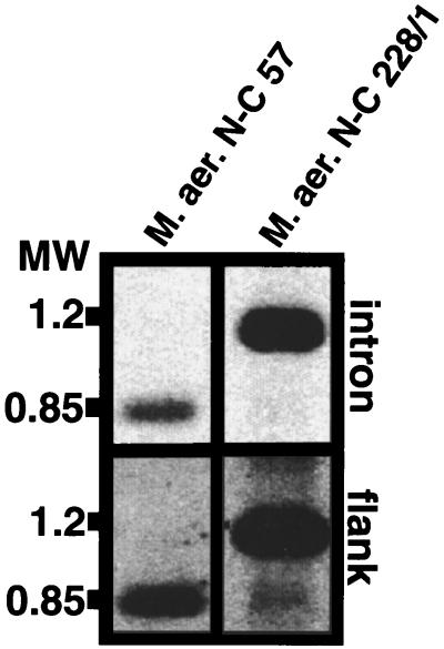
The origin and evolution of bacterial introns are still controversial issues. Here we present data on the distribution and evolution of a recently discovered divergent tRNA(Leu)(UAA) intron. The intron shows a higher sequence affiliation with introns in tRNA(Ile)(CAU) and tRNA(Arg)(CCU) genes in alpha- and beta-proteobacteria, respectively, than with other cyanobacterial tRNA(Leu)(UAA) group I introns. The divergent tRNA(Leu)(UAA) intron is sporadically distributed both within the Nostoc and the Microcystis radiations. The complete tRNA gene, including flanking regions and intron from Microcystis aeruginosa strain NIVA-CYA 57, was sequenced in order to elucidate the evolutionary pattern of this intron. Phylogenetic reconstruction gave statistical evidence for different phylogenies for the intron and exon sequences, supporting an evolutionary model involving horizontal intron transfer. The distribution of the tRNA gene, its flanking regions, and the introns were addressed by Southern hybridization and PCR amplification. The tRNA gene, including the flanking regions, were absent in the intronless stains but present in the intron-containing strains. This suggests that the sporadic distribution of this intron within the Microcystis genus cannot be attributed to intron mobility but rather to an instability of the entire tRNA(Leu)(UAA) intron-containing genome region. Taken together, the complete data set for the evolution of this intron can best be explained by a model involving a nested evolution of the intron, i.e., wherein the intron has been transferred horizontally (probably through a single or a few events) to a tRNA(Leu)(UAA) gene which is located within a unstable genome region.


Figures

FIG. 1.
FIG. 1.
Cloned flanking region, included probe, and primer regions for the primers used to generate the probes for M. aeruginosa N-C 57 (EMBL accession no. AJ307004). Two partial ORFs (RF1 and RF2) were identified in the flanking region. The primer sites and orientation are indicated with arrows, while the probe regions are indicated with bars. The type B tRNALeu(UAA) exon probe is denoted with a bar with a stippled line to indicate that stippled region is not present in the probe.
FIG. 2.
FIG. 2.
Secondary structure of the intron-containing tRNALeu(UAA) gene. The intron insertion site is indicated by an arrow. The structure was obtained by a combination of MFOLD computer prediction (GCG Package; Genetic Computer Group, Madison, Wis.) and manual editing.
FIG. 3.
FIG. 3.
Phylogenetic reconstruction of tRNA exon (A) and intron (B) sequences. The trees were built by using both distance and maximum-parsimony methods. The trees shown in panels A and B (based on 100 and 275 aligned positions, respectively) were built by the neighbor-joining method using maximum-likelihood distances. The distances are expressed as substitutions per nucleotide in the neighbor-joining tree. Numbers at the nodes (expressed as maximum likelihood/LogDet distances/maximum-likelihood distances/heuristic search for maximum parsimony trees) indicate the percentage of Puzzle (for maximum likelihood) and bootstrap trees for the others in which the cluster descending from the node was found. Numbers are only shown for nodes supported by ≥50% for all of the methods tested. Each tRNA gene is annotated in panel A, while the three clusters of introns identified (panel B) are indicated by I, II, and III.
FIG. 4.
FIG. 4.
PCR amplification analyses with the primer pair CAB-B-CBB (A) and through control amplification of 16S rDNA with the primer pair CC-CD (B). All samples were electrophoresed in 1.5% agarose gels for 30 min at 100 V. Twenty percent of the amplification products was loaded in each lane. MW, molecular weight marker.
FIG. 5.
FIG. 5.
Southern hybridization analysis of selected Microcystis strains. Genomic DNA digested with HindIII was separated, blotted, and hybridized as described in Materials and Methods. The same membrane was used in each of the hybridizing experiments, providing an exact assignment of the hybridizing bands from different experiments. The hybridizing probes were as follows: flank 1 (A), tRNA type A (B), tRNA type B (C), intron (D), and 16S rDNA (E).
FIG. 6.
FIG. 6.
Southern hybridization analysis with AluI-digested DNA for the M. aeruginosa strains N-C 57 and 228/1. Digested genomic DNA was separated, blotted, and hybridized as described in Materials and Methods. The following probes were used: flank 2 (A) and intron (B).
FIG. 7.
FIG. 7.
Model for the evolution of the Microcystis tRNALeu(UAA) group I intron. The model shows that the process of intron homing is relatively infrequent (A) and that the process of recombination of the entire tRNALeu(UAA) genome region is relatively frequent (B). Taken together, these two processes can explain the origin of the Microcystis introns and the current intron distribution within this genus.

Similar articles

Cited by

References

    1. Belfort, M., M. E. Reaban, T. Coetzee, and J. Z. Dalgaard. 1995. Prokaryotic introns and inteins: a panoply of form and function. J. Bacteriol. 177:3897–3903. - PMC - PubMed
    1. Besendahl, A., Y. L. Qiu, J. Lee, J. D. Palmer, and D. Bhattacharya. 2000. The cyanobacterial origin and vertical transmission of the plastid tRNA(Leu) group-I intron. Curr. Genet. 37:12–23. - PubMed
    1. Bonocora, R. P., and D. A. Shub. 2001. A novel group I intron-encoded endonuclease specific for the anticodon region of tRNA fMet genes. Mol. Microbiol. 39:1299–1306. - PubMed
    1. Castenholz, R. W., and J. B. Waterbury. 1989. Group I. Cyanobacteria, p.1710–1728. In N. Pfenning and J. G. Holt (ed.), Bergey’s manual of systematic bacteriology. The Williams & Wilkins Co., Baltimore, Md.
    1. Edgell, D. R., M. Belfort, and D. A. Shub. 2000. Barriers to intron promiscuity in bacteria. J. Bacteriol. 182:5281–5289. - PMC - PubMed

Publication types

Associated data

LinkOut - more resources