Comparative Study
. 2002 Feb;184(3):687-94.
doi: 10.1128/JB.184.3.687-694.2002.

Three proliferating cell nuclear antigen-like proteins found in the hyperthermophilic archaeon Aeropyrum pernix: interactions with the two DNA polymerases

Affiliations
Comparative Study

Three proliferating cell nuclear antigen-like proteins found in the hyperthermophilic archaeon Aeropyrum pernix: interactions with the two DNA polymerases

Katsuya Daimon et al. J Bacteriol. 2002 Feb.

Abstract

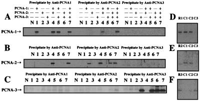
Proliferating cell nuclear antigen (PCNA) is an essential component in the eukaryotic DNA replication machinery, in which it works for tethering DNA polymerases on the DNA template to accomplish processive DNA synthesis. The PCNA also interacts with many other proteins in important cellular processes, including cell cycle control, DNA repair, and an apoptotic pathway in the domain EUCARYA: We identified three genes encoding PCNA-like sequences in the genome of Aeropyrum pernix, a crenarchaeal archaeon. We cloned and expressed these genes in Escherichia coli and analyzed the gene products. All three PCNA homologs stimulated the primer extension activities of the two DNA polymerases, polymerase I (Pol I) and Pol II, identified in A. pernix to various extents, among which A. pernix PCNA 3 (ApePCNA3) provided a most remarkable effect on both Pol I and Pol II. The three proteins were confirmed to exist in the A. pernix cells. These results suggest that the three PCNAs work as the processivity factor of DNA polymerases in A. pernix cells under different conditions. In Eucarya, three checkpoint proteins, Hus1, Rad1, and Rad9, have been proposed to form a PCNA-like ring structure and may work as a sliding clamp for the translesion DNA polymerases. Therefore, it is very interesting that three active PCNAs were found in one archaeal cell. Further analyses are necessary to determine whether each PCNA has specific roles, and moreover, how they reveal different functions in the cells.


Figures

FIG. 1.
FIG. 1.
Purification of recombinant ApePCNA homologs from E. coli cells. All samples of each step were fractionated on SDS-10% polyacrylamide gels, which were stained with Coomassie brilliant blue. The sizes of the molecular mass markers (Protein Marker, Broad Range; New England BioLabs, Inc.) are indicated on the left of each figure. The arrows on the right side of the panels show the obtained proteins. (A) Purification steps of recombinant ApePCNA1. Lanes: M, molecular mass markers; 1, crude cell extract after sonication; 2, supernatant from heat treatment (75°C, 15 min.); 3, supernatant after polyethyleneimine treatment; 4, HiTrap Q fraction; and 5, HiTrap heparin fraction. (B) Purification steps of recombinant ApePCNA2. Lanes: M, molecular mass markers; 1, crude cell extract after sonication; 2, supernatant after polyethyleneimine treatment; 3, HiTrap Phenyl Sepharose HP fraction; 4, HiTrap Q fraction; 5, and HiTrap heparin fraction. (C) Purification steps of recombinant ApePCNA3. All lanes are the same as in the case of ApePCNA1.
FIG. 2.
FIG. 2.
Identification of ApePCNAs in A. pernix cells. (A) Recombinant ApePCNA1 (lane 1, 40 ng) and A. pernix cell extracts (lane 2, 30 μg) were separated by SDS-10% PAGE and then were analyzed by Western blotting with anti-ApePCNA1 antiserum. (B) Recombinant ApePCNA2 (lane 1, 80 ng) and A. pernix cell extracts (lane 2, 30 μg) were analyzed with anti-ApePCNA2 antiserum. (C) Recombinant ApePCNA3 (lane 1, 10 ng) and A. pernix cell extracts (lane 2, 15 μg) were analyzed with anti-ApePCNA3 antiserum.
FIG. 3.
FIG. 3.
Detection of the reaction products by Pol I and Pol II. The primer elongation abilities of Pol I and Pol II in the absence and presence (0.3 μg) of ApePCNA1 (lane 1), ApePCNA2 (lane 2), and ApePCNA3 (lane 3) were investigated by using two templates: circular M13 ssDNA (panel A) and linearized M13 ssDNA (panel B). Lanes N and P indicate reactions without PCNA and with PfuPCNA, respectively, in both panels. The products were analyzed by 1.2% alkaline agarose gel electrophoresis, and the reaction products were visualized by autoradiography. The sizes indicated on the right side were from BstPI-digested lambda DNA labeled by polynucleotide kinase with [γ-32P]ATP. The arrow shows the position of the product with full-length M13 DNA.
FIG. 4.
FIG. 4.
Association states of ApePCNAs assessed by chemical cross-linking. ApePCNA1 (A), ApePCNA2 (B), and ApePCNA3 (C) were treated with buffer (lane1) and sulfo-EGS (lane 2) for 2 min and were analyzed by SDS-10% PAGE, followed by Western blotting using anti-ApePCNA1, anti-ApePCNA2, and anti-ApePCNA3, respectively. Lanes 3 in panel A and B include equal amounts of ApePCNA1 and ApePCNA2. The cross-linked products from the mixture of ApePCNA1 and ApePCNA2 are indicated by a thick arrow. Each band is derived from a PCNA monomer (a), a single cross-linked dimer (b), three cross-linked circled trimers (c), and two cross-linked linear trimers (d), as indicated for the T4 gp45 protein (19). The indicated molecular sizes were derived from the prestained protein markers (APRO Science, Inc.).
FIG. 5.
FIG. 5.
Analysis of heterologous interactions of ApePCNAs. (A to C) Immunoprecipitation analysis was done using purified ApePCNAs and anti-ApePCNA1 (left part of each panel), anti-ApePCNA2 (middle part), and anti-ApePCNA3 (right part). The immunocomplexes were captured with protein A-Sepharose and were subjected to SDS-12% PAGE, followed by Western blot analysis using anti-PCNA1 (A), anti-PCNA2 (B), and anti-PCNA3 (C). Lanes: N, no protein; 1, ApePCNA1; 2, ApePCNA2; 3, ApePCNA3; 4, ApePCNA1 + ApePCNA2; 5, ApePCNA1 + ApePCNA3; 6, ApePCNA2 + ApePCNA3; and 7, ApePCNA1 + ApePCNA2 + ApePCNA3. (D to F) Total cell extract of A. pernix was reacted with anti-ApePCNA1 (lane C1), anti-ApePCNA2 (lane C2), and anti-ApePCNA3 (lane C3). These complexes were captured with protein A-Sepharose, and the immunoprecipitates were analyzed as described above, using anti-ApePCNA1 (D), anti-ApePCNA2 (E), and anti-ApePCNA3 (F). As a positive control, purified ApePCNAs precipitated by anti-PCNAs were loaded onto each gel (lanes: R1, ApePCNA1; R2, ApePCNA2; and R3, ApePCNA3).
FIG. 6.
FIG. 6.
Amino acid sequence alignment of the three ApePCNAs, P. furiosus, yeast, and human PCNAs. Identical and similar amino acid residues are boxed with black and gray, respectively. Important regions required for the function and oligomerization of PCNA, predicted from previous studies, are indicated with the alignment. Ape, A. pernix; Pfu, P. furiosus; Sce, Saccharomyces cerevisiae; Hsa, Homo sapiens.

Similar articles

Cited by

References

    1. Bocquier, A. A., L. Liu, I. K. O. Cann, K. Komori, D. Kohda, and Y. Ishino. 2001. Archaeal primase: bridging the gap between RNA and DNA polymerase. Curr. Biol. 11:452–456. - PubMed
    1. Cai, R. L., Y. Yan-Neale, M. A. Cueto, H. Xu, and D. Cohen. 2000. HDAC1, a histone deacetylase, forms a complex with Hus1 and Rad9, two G2/M checkpoint Rad proteins. J. Biol. Chem. 275:27909–27916. - PubMed
    1. Cann, I. K. O., K. Komori, H. Toh, S. Kanai, and Y. Ishino. 1998. A heterodimeric DNA polymerase: evidence that members of Euryarchaeota possess a distinct DNA polymerase. Proc. Natl. Acad. Sci. USA 95:14250–14255. - PMC - PubMed
    1. Cann, I. K. O., S. Ishino, N. Nomura, Y. Sako, and Y. Ishino. 1999. Two family B DNA polymerases from Aeropyrum pernix, an aerobic hyperthermophilic crenarchaeote. J. Bacteriol. 181:5984–5992. - PMC - PubMed
    1. Cann, I. K. O., and Y. Ishino. 1999. Archaeal DNA replication: identifying the pieces to solve a puzzle. Genetics 152:1249–1267. - PMC - PubMed

Publication types

MeSH terms