. 2002 Jan 21;156(2):299-313.
doi: 10.1083/jcb.200109037. Epub 2002 Jan 14.

Ras and TGF[beta] cooperatively regulate epithelial cell plasticity and metastasis: dissection of Ras signaling pathways

Affiliations

Ras and TGF[beta] cooperatively regulate epithelial cell plasticity and metastasis: dissection of Ras signaling pathways

Elzbieta Janda et al. J Cell Biol. .

Abstract

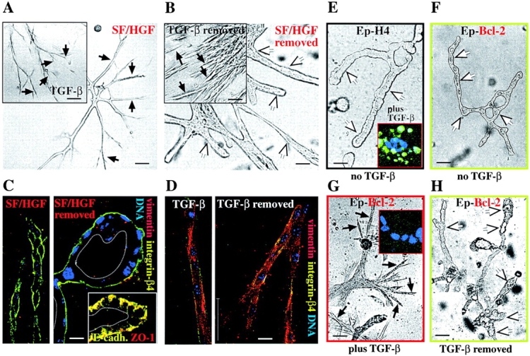
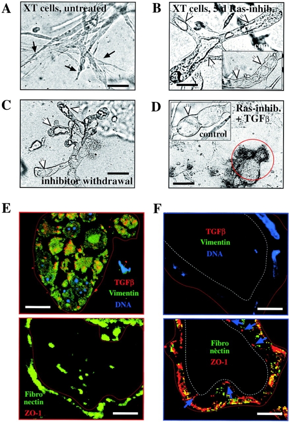
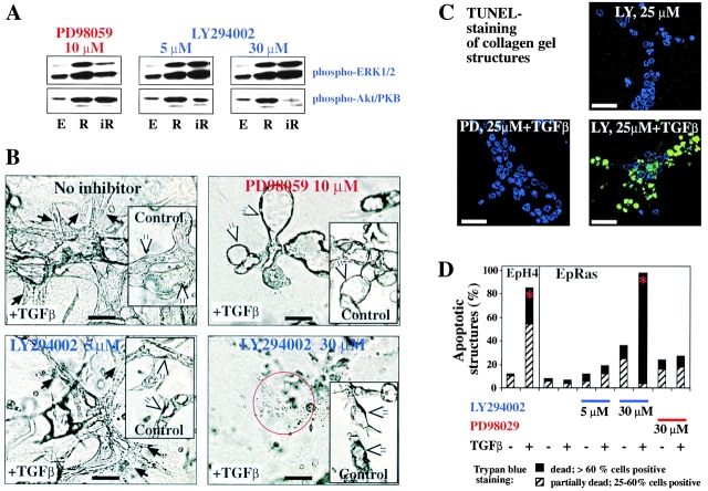
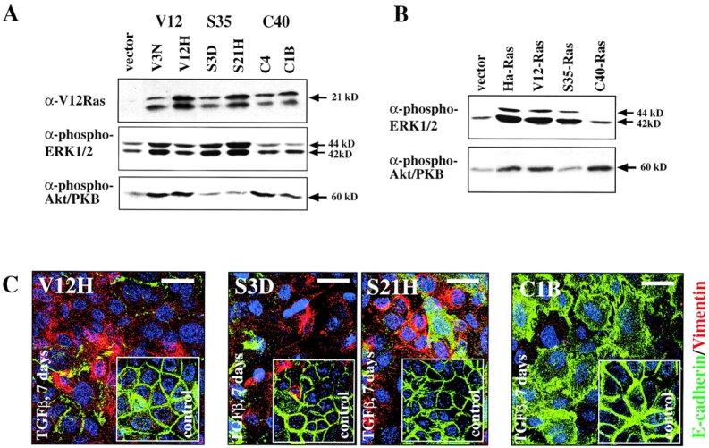
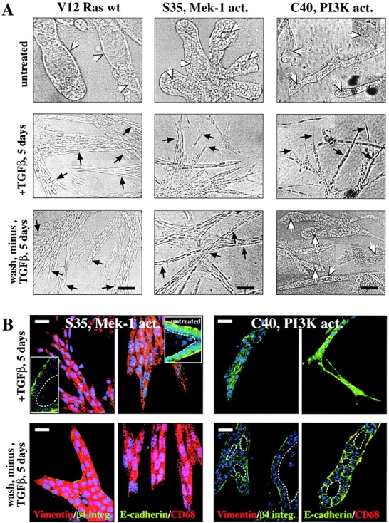
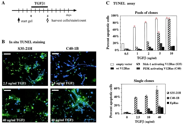
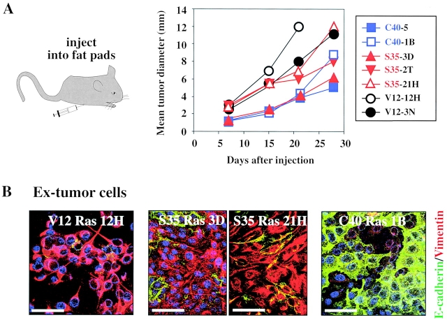
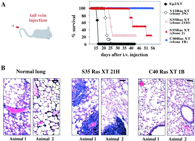
Multistep carcinogenesis involves more than six discrete events also important in normal development and cell behavior. Of these, local invasion and metastasis cause most cancer deaths but are the least well understood molecularly. We employed a combined in vitro/in vivo carcinogenesis model, that is, polarized Ha-Ras-transformed mammary epithelial cells (EpRas), to dissect the role of Ras downstream signaling pathways in epithelial cell plasticity, tumorigenesis, and metastasis. Ha-Ras cooperates with transforming growth factor beta (TGFbeta) to cause epithelial mesenchymal transition (EMT) characterized by spindle-like cell morphology, loss of epithelial markers, and induction of mesenchymal markers. EMT requires continuous TGFbeta receptor (TGFbeta-R) and oncogenic Ras signaling and is stabilized by autocrine TGFbeta production. In contrast, fibroblast growth factors, hepatocyte growth factor/scatter factor, or TGFbeta alone induce scattering, a spindle-like cell phenotype fully reversible after factor withdrawal, which does not involve sustained marker changes. Using specific inhibitors and effector-specific Ras mutants, we show that a hyperactive Raf/mitogen-activated protein kinase (MAPK) is required for EMT, whereas activation of phosphatidylinositol 3-kinase (PI3K) causes scattering and protects from TGFbeta-induced apoptosis. Hyperactivation of the PI3K pathway or the Raf/MAPK pathway are sufficient for tumorigenesis, whereas EMT in vivo and metastasis required a hyperactive Raf/MAPK pathway. Thus, EMT seems to be a close in vitro correlate of metastasis, both requiring synergism between TGFbeta-R and Raf/MAPK signaling.


Figures

Figure 1.
Figure 1.
Scattering induced by scatter factor (HGF/SF) and TGFβ can be distinguished from EMT by reversibility and lack of marker changes. (A–D) EpRas collagen gel structures were treated with SF/HGF and TGFβ. (A) EpRas cells seeded in collagen gels for 3 d were treated with HGF/SF or TGFβ (insets) for an additional 5 d and photographed (B). After factor removal, (as described in Materials and methods) cells were maintained for an additional 4 d and photographed again. White arrows indicate lumina of tubular structures; black arrows indicate unordered strands of spindle-like invasive cells. (C and D). Structures treated as in A and B were fixed, stained with antibodies to the epithelial markers β4-integrin, E-cadherin, and ZO-1 (C, insets) and the mesenchymal marker vimentin, and analyzed by confocal immunofluorescence microscopy (as described in Materials and methods). (C) Structures transiently treated with HGF/SF. (D) Structures transiently treated with TGFβ. Note that HGF/SF-treated cells retain nonpolar distributed β4-integrin (C, left), whereas cells completely repolarize after HGF/SF removal (basolateral localized E-cadherin, and ZO-1) (C, left and inset). In contrast, the TGFβ-treated control structures undergo EMT (loss of β4-integrin, and vimentin expression) (D, left), persisting after TGFβ withdrawal (D, right). (E–H) Bcl-2–expressing EpH4 cells (Ep–Bcl-2) and control EpH4 cells were treated with TGFβ, which induced apoptosis (revealed by in situ TUNEL staining) in the EpH4 cells (E, inset) but a spindle cell-like morphology (G) and no apoptosis in the Ep–Bcl-2 cells (G, inset). Untreated cells of both types form normal hollow tubules (E and F). After withdrawal of TGFβ, the EpH4–Bcl-2 cells revert to tubular structures (H). Bars: (A, B, and E–H) 50 μm; (C and D) 10 μm.
Figure 2.
Figure 2.
Ras activity is required for induction and maintenance of EMT. (A–D) EpXT cells were seeded into collagen gels in the absence (A) or presence (B) of 10 μM Ras farnesylation inhibitor (L739749), and resulting structures were photographed after 6 d (as described in Materials and methods). Note the complete reversal of EpXT cells to tubular structures with lumina (white arrows), which persisted after removal of the inhibitor for 4 d (C). The inhibitor alone did not affect tubular structures formed by EpRas cells (B, inset) or after reversal (D, inset, control), whereas treatment of the reverted structures with TGFβ plus L739749 caused cell disintegration. (E and F) Immunostaining of frozen ultrathin sections of EpXT collagen gel structures before (E) or after treatment (F) with L739749 for 5 d. Sections were stained with antibodies to TGFβ (red) (Oft et al., 1996) and vimentin (green) plus DAPI (top, DNA). Parallel sections (bottom) were stained with antibodies to ZO-1 (red) and fibronectin (green). L739749 causes loss of intracellular TGFβ and vimentin (top) and extracellular fibronectin (bottom) and reexpression of ZO-1 at apicolateral sites of tight junctions (blue arrows, bottom left). Bars: (A–D) 50 μm; (E and F) 20 μm.
Figure 3.
Figure 3.
Inhibitors of Mek-1 prevent and reverse EMT, and PI3K inhibitors abolish protection from TGFβ-induced apoptosis. (A) EpH4 cells (lanes marked E) or EpRas cells mock treated (lanes marked R) or treated with the inhibitors indicated (lanes marked iR) were lysed after 6 h of inhibitor treatment and analyzed for serine phosphorylation of Erk1/2 (MAPK) and PKB/Akt in Western blots (as described in Materials and methods). (B) EpRas cells seeded into collagen gels in the presence or absence (insets) of 5 ng/ml TGFβ. Both types of cultures were then left untreated (top, control) or treated with PD98059 (top right) and LY294002 (bottom) for another 5 d (as described in Materials and methods). Neither inhibitor was toxic to EpRas cells without TGFβ (insets; LY294002 slowed down structure growth). PD98059 (10 μM) completely reverted EMT (white arrows, top right), 5 μM LY294002 had no effect on the mesenchymal structures (black arrows, bottom left), whereas 30 μM caused cell death (bottom right, red circle). (C) Confocal analysis of in situ TUNEL staining (green) performed on collagen gel structures from EpRas cells treated for 4 d as indicated on the microphotographs (counterstaining for DNA in blue). (D) Quantitative analysis of cell death caused by inhibition of MEK-1 and PI3K in the absence or presence of TGFβ using trypan blue dye exclusion assay. The percentages of structures consisting of 25–60% (hatched bars, partially dead) or >60% trypan blue–positive cells (black bars, dead) are shown (as described in Materials and methods). Red asterisks indicate statistically significant induction of apoptosis by TGFβ. Bars: (B and C) 50 μm.
Figure 4.
Figure 4.
Biochemical and biological characterization of EpH4 cells expressing Ras effector-specific mutants lacking signaling along the MAPK or PI3K pathways. (A) Clones of EpH4 cells expressing the Ras effector mutants S35-Ras, C40-Ras, or V12-Ras were tested for overexpression of Ras proteins in Western blots using V12-Ras–specific antibodies. Clones highly overexpressing the exogenous Ras proteins (top) were then tested for levels of phospho-Erk (middle) or phospho-Akt (bottom) by respective phospho-specific antibodies (as described in Materials and methods). (B) Mass cultures (pools of multiple clones) expressing lower but still clearly elevated levels of H-V12–Ras or Ras effector mutant proteins S35 or C40 (unpublished data) were analyzed for phospho-Erk and phospho-Akt expression as in A. (C) Representative clones expressing V12-Ras (left), S35-Ras (middle), and C40-Ras (right) were cultivated on porous supports for 7 d in the presence (panels) or absence of TGFβ (insets). Cells were immunostained for E-cadherin (green) and vimentin (red) plus DAPI counterstaining for DNA (blue; as described in Materials and methods) and analyzed by confocal immunofluorescence microscopy. Bars, 20 μm.
Figure 5.
Figure 5.
EpH4 cells expressing V12-Ras or S35-Ras undergo EMT upon TGFβ treatment; C40-Ras cells undergo scattering. (A) EpH4 cell clones overexpressing V12-Ras (left) and the Ras effector mutants S35-Ras (middle) and C40-Ras (right; Fig. 4) were seeded in collagen gels and either left untreated (top) or treated (middle) with 5 ng/ml TGFβ for 5 d followed by removal of TGFβ and further cultivation for 5 d (bottom). Photographs of representative tubular structures with lumina (white arrows) or distended chords and strands of invasive cells with mesenchymal morphology (black arrows) are shown. (B) Similar collagen gel structures as in A were stained in situ with antibodies to epithelial markers (green, β4-integrin and E-cadherin) and mesenchymal markers (red, vimentin and CD68; as described in the text) and analyzed by confocal immunofluorescence microscopy (as described in Materials and methods). (Top left, insets) tubular structures formed by untreated S35-Ras cells. (Top) “Invasive” structures formed by S35-Ras and C40-Ras cells after TGFβ treatment (note the persistent nonpolarized expression of epithelial markers in the C40-Ras structures). (Bottom) Structures formed by S35-Ras and C40-Ras after removal of TGFβ (mesenchymal shape and mesenchymal markers persist in the S35-Ras cells; C40-Ras cells revert to tubular structures with lumina [white dotted lines] basolaterally expressed epithelial markers). Bars: (A) 50 μm; (B) 20 μm.
Figure 6.
Figure 6.
V12-Ras and C40-Ras but not S35-Ras protect EpH4 cells from TGFβ-induced apoptosis. (A) For apoptosis determination in collagen gels, EpH4 cells expressing Ras or Ras effector–specific mutants were allowed to form structures in collagen gels for 3 d and were treated with various concentrations of TGFβ (0.5–40 ng/ml) for 4 d (dotted bar). Cells were either TUNEL stained in suspension (C, top) or in situ (B, and C, bottom panel; as described in Materials and methods). (B) Confocal immunofluorescence analysis of gel structures formed by clones overexpressing S35-Ras (left) and C40-Ras cells (right) treated with moderate (top, 2.5 ng/ml) or high levels of TGFβ (40 ng/ml; see A) and subjected to in situ TUNEL staining (green). DAPI staining (blue) indicates live cells. (C) Quantitation of TUNEL-positive cells from collagen gel structures either stained and counted in suspension after collagenase digestion (top; red asterisks indicate statistically significant increases in apoptosis induced by TGFβ) or stained and counted in situ (bottom; as described in Materials and methods). Mean percentages of apoptotic cells from multiple gel structures plus standard deviations (error bars) from three independent determinations are shown. Bars, 50 μm.
Figure 7.
Figure 7.
S35-Ras– and C40-Ras–overexpressing cells form tumors, but only S35-Ras cells undergo EMT in vivo. (A) Various clones (see also Fig. 4 A) overexpressing V12-Ras (black symbols) S35-Ras (red symbols) and C40-Ras (blue symbols) were injected into the mammary gland fat pads of mice (four mice with a total of 16 injection sites per clone). Growth of the multiple tumors developing in all mice was followed by measuring mean tumor sizes (as described in Materials and methods). (B) Ex-tumor cells recultivated from 4–5-wk-old tumors (as described in Materials and methods) induced by V12-Ras, S35-Ras, and C40-Ras clones (A) were seeded onto porous supports and analyzed by immunostaining for E-cadherin (green) and vimentin (red). Blue, DAPI counterstain for DNA. Bars, 20 μm.
Figure 8.
Figure 8.
S35-Ras– but not C40-Ras–overexpressing EpH4 cells form lung metastases upon tail vein injection. (A) Ex-tumor cells from the V12-Ras (black symbols), S35-Ras (red symbols), and C40-Ras clones (blue symbols) indicated were injected intravenously into four nude mice per cell type. Moribund mice were killed and analyzed for the presence of lung metastases. The two surviving mice injected with S35-Ras clones were killed at day 55 and also contained lung metastases. None of the four mice injected with C40-Ras cells (killed after 10 wk) showed any lung metastases. (B) Histological stainings of sections from lungs of uninjected animals (left) and S35-Ras (middle) and C40-Ras–injected mice (two animals each). Note large metastases (also around blood vessels) only in the S35-Ras–injected mice. Bar, 100 μm.

References

    1. Bakin, A.V., A.K. Tomlinson, N.A. Bhowmick, H.A. Moses, and C.L. Arteaga. 2000. Phosphatidylinositol 3-kinase function is required for transforming growth factor β-mediated epithelial to mesenchymal transition and cell migration. J. Biol. Chem. 275:36803–36810. - PubMed
    1. Bergers, G., and L.M. Coussens. 2000. Extrinsic regulators of epithelial tumor progression: metalloproteinases. Curr. Opin. Genet. Dev. 10:120–127. - PubMed
    1. Boyer, B., A.M. Valles, and J.P. Thiery. 1996. Model systems of epithelium-mesenchyme transitions. Acta Anat. 156:227–239. - PubMed
    1. Boyer, B., A.M. Valles, and N. Edme. 2000. Induction and regulation of epithelial-mesenchymal transitions. Biochem. Pharmacol. 60:1091–1099. - PubMed
    1. Brinkmann, V., H. Foroutan, M. Sachs, K.M. Weidner, and W. Birchmeier. 1995. Hepatocyte growth factor/scatter factor induces a variety of tissue-specific morphogenic programs in epithelial cells. J. Cell Biol. 131:1573–1586. - PMC - PubMed

Publication types

MeSH terms

Substances