. 2002 Jan 15;538(Pt 2):343-55.
doi: 10.1113/jphysiol.2001.012839.

Stimulation of recombinant Ca(v)3.2, T-type, Ca(2+) channel currents by CaMKIIgamma(C)

Affiliations

Stimulation of recombinant Ca(v)3.2, T-type, Ca(2+) channel currents by CaMKIIgamma(C)

Joshua T Wolfe et al. J Physiol. .

Abstract

Molecular cloning of low-voltage activated (LVA) T-type calcium channels has enabled the study of their regulation in heterologous expression systems. Here we investigate the regulation of Ca(v)3.2 alpha(1)-subunits (alpha1H) by calcium- and/or calmodulin-dependent protein kinase II (CaMKII). 293 cells stably expressing alpha1H were transiently transfected with CaMKIIgamma(C). Using the whole-cell recording configuration, we observed that elevation of pipette free Ca(2+) (1 microM) in the presence of CaM (2 microM) increases T-type channel activity selectively at negative potentials, evoking an 11 mV hyperpolarizing shift in the half-maximal potential (V(1/2)) for activation. The V(1/2) of channel inactivation is not altered by Ca(2+)/CaM. These effects reproduced modulation observed in adrenal zona glomerulosa cells. The potentiation by Ca(2+)/CaM was dependent on the co-expression of CaMKIIgamma(C) and required Ca(2+)/CaM-dependent kinase activity. Peptide (AIP) and lipophilic (KN-62) protein kinase inhibitors prevented the Ca(2+)/CaM-induced changes in channel gating without altering basal Ca(v)3.2 channel activity (27 nM free Ca(2+)) as did replacing pipette ATP with adenylyl imidodiphosphate (AMP-PNP), a non-hydrolysable analogue. CaMKII-dependent potentiation of channel opening resulted in significant increases in apparent steady-state open probability (P(o)) and sustained channel current at negative voltages. Under identical conditions, CaMKII activation did not regulate the activity of Ca(v)3.1 channels, the first cloned member (alpha1G) of the T-type Ca(2+) channel family. Our results provide the first evidence for the differential regulation of two members of the Ca(v)3 family by protein kinase activation and the first report reconstituting CaMKII-dependent regulation of any cloned Ca(2+) channel.


Figures
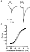
Figure 1
Figure 1. Elevation of pipette Ca2+/CaM fails to increase Ca2+ currents in 293 cells expressing Cav3.2 channels
Slowly deactivating tail currents were elicited at −60 mV after various depolarizing test pulses (Vt = −60 to +10 mV; 10 ms) from a holding potential of −90 mV. A, sample currents at Vt = −30 and +10 mV from two cells with intracellular free Ca2+ fixed at 27 nm Ca2+ (○) or 1 μm Ca2+ + 2 μm CaM (•). Note that the two cells have similar maximal currents (Imax). B, voltage dependence of activation. Normalized amplitude of slowly deactivating tail current (I/Imax) plotted (means ± s.e.m.) vs. Vt for two data sets. Data were fitted to a squared Boltzmann distribution yielding half-maximal activation potentials of: 27 nm Ca2+, V1/2 = −23.7 ± 0.4 mV (k = 10.2, r = 0.99; n = 14 cells) and 1 μm Ca2+, V1/2 = −26.8 ± 0.2 mV * (k = 10.2, r = 0.99, n = 9 cells, where k is the slope factor and r is the regression coefficient. *Not statistically different from 27 nm Ca2+ (by Student' unpaired t test).
Figure 2
Figure 2. Identification of two partial CaMKIIγC transcripts in bovine glomerulosa cells
Deduced amino acid sequences from two bovine glomerulosa cell RT-PCR products (γC and γX) amplified using bovine glomerulosa cell RNA and primers to highly conserved regions in the regulatory and association domains in all CaMKII gene families that spanned the intervening variable regions. The bovine sequences were aligned showing deletion regions (V1 and V2) absent in CaMKIIγ subunits previously designated as γC. The conserved domain separating the V1 and V2 regions was absent in γX. The sequences have been deposited in GenBank with the following accession numbers: bγC, AF 389986; and bγX, AF389987.
Figure 3
Figure 3. Elevation of pipette Ca2+/CaM induces a hyperpolarizing shift in the V1/2 of activation of Cav3.2 channels in 293 cells expressing CaMKIIγC
Slowly deactivating tail currents were elicited at −60 mV, after various depolarizing test pulses (Vt = −60 to +10 mV; 10 ms) from a holding potential of −90 mV (activation) or after depolarizations (+20 mV, 8 ms) from various prepulse potentials (VP = −90 to −20 mV) lasting 6 s (inactivation). A, activation. Sample currents at Vt = −55, −45, −35, −25, −5 or +5 mV from two cells with intracellular free Ca2+ fixed at 27 nm (○) or 1 μm + 2 μm CaM (•). Note the larger amplitude of currents at hyperpolarized potentials recorded with free Ca2+ fixed at 1 μm (•). B, voltage dependence of channel activation. Relative amplitude of tail currents (means ± s.e.m.) plotted vs. Vt for two data sets. Data were fitted to a squared Boltzmann distribution, yielding half-maximal activation potentials of: 27 nm Ca2+, V1/2 = −23.1 ± 0.3 mV (k = 11.2, r = 0.99; n = 21 cells); and 1 μm Ca2+, V1/2 = −34.0 ± 0.5 mV * (k = 10.2, r = 0.99, n = 21 cells). * Statistically different from 27 nm Ca2+ (P < 0.05 by Mann-Whitney unpaired non-parametric test). Inset, immunoblot of cell lysates from Cav3.2-expressing cells transfected with: CaMKIIγC and GFP (γC transfected), or GFP alone (untransfected). Recombinant CaMKIIγC protein served as standard. C, inactivation. Sample currents at VP = −90, −70, −60, −55, −50 and −40 mV from two cells with intracellular free Ca2+ fixed at 27 nm (▵) or 1 μm + 2 μm CaM (▾). D, voltage dependence of channel inactivation. Relative amplitude of tail current (means ± s.e.m.) plotted vs. Vp for two data sets. Data were fitted to a Boltzmann distribution, yielding half-maximal inactivation potentials of: 27 nm Ca2+, V1/2 = −67.1 ± 0.2 mV (k = 6.6, r = 0.99, n = 10 cells); and 1 μm Ca2+, V1/2 = −67.2 ± 0.2 mV * (k = 6.8, r = 0.99, n = 9 cells). * Not statistically significant from 27 nm Ca2+ (by Student' unpaired t test).
Figure 4
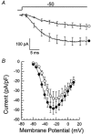
Figure 4. Ca2+/CaM shifts the current-voltage relationship of Cav3.2 channels to negative potentials
A, mean traces (n = 10 and 11) at Vt = −50 mV show inward currents elicited from cells with pipette Ca2+ fixed at 27 nm (○) or 1 μm (•). B, I-V relationship. Peak inward current normalized to cell capacitance plotted vs. test potential for cells recorded with CaMKII-activating (n = 11 cells) or non-activating solutions (n = 10 cells). Note that with CaMKII-activating solutions Cav3.2 currents are enhanced within the range of −60 to −35 mV.
Figure 5
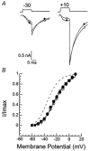
Figure 5. Autocamtide-2-related inhibitory protein (AIP), a specific peptide inhibitor of CaMKII, blocks Ca2+/CaM-induced potentiation of Cav3.2 currents in 293 cells expressing CaMKIIγC
A, representative traces at Vt = −30 mV and +10 mV show tail currents (Vr= −60 mV) elicited from two cells with 2 μm AIP in the pipette solution. Intracellular free Ca2+ was fixed at 27 nm (○) or 1 μm (•). Note that in the presence of AIP, Ca2+/CaM did not potentiate Cav3.2 currents. B, voltage dependence. Relative amplitude of tail current (means ± s.e.m.) plotted vs. Vt for two data sets. Half-maximal activation potentials were calculated as in Fig. 3B; 27 nm Ca2+ + AIP, V1/2 = −21.5 ± 0.3 mV (k = 12.4, r = 0.99, n = 12 cells); and 1 μm Ca2+ + AIP, V1/2 = −22.7 ± 0.5 mV * (k = 12.4, r = 0.99, n = 11 cells). * Not statistically significant from 27 nm Ca2+ + AIP (by Student' unpaired t test). Dashed lines show comparison fits to data points obtained at 27 nm and 1 μm Ca2+ without AIP in the pipette, data from Fig. 3B.
Figure 6
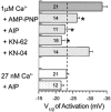
Figure 6. CaMKII activity is required for Ca2+/CaM to modify the gating of Cav3.2 channels
V1/2 of activation (means ± s.e.m., n = number of cells) was calculated for each cell recorded with 1 μm or 27 nm Ca2+ in the absence and presence of kinase inhibitors in the bath or pipette solutions. Dashed line indicates baseline V1/2 determined in Fig. 3B at 27 nm Ca2+ in the absence of inhibitors. * Statistically significant from 27 nm Ca2+ (P < 0.05 by non-parametric one-way ANOVA). AMP-PNP (3 μm), poorly hydrolysable ATP analogue; AIP (2 μm), specific CaMKII-peptide inhibitor; and KN-62 (3 μm), membrane-permeable CaMKII-inhibitor, significantly reduced Ca2+-induced gating change. KN-04 (3 μm), inactive CaMKII-inhibitor, preserved gating change. Neither AIP nor KN-04 (not shown) changed control V1/2 of activation at 27 nm Ca2+.
Figure 7
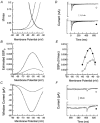
Figure 7. CaMKII activation increases Cav3.2 window current
A, predicted voltage range for observable Cav3.2 window currents. Overlap of Boltzmann distributions describing the voltage dependence of Cav3.2 activation (m2) and inactivation (h) determined from data in Fig. 3 defines voltage ranges for discernible window currents. Data were obtained with non-activating (continuous lines) or CaMKII-activating (dashed lines) pipette solutions. Note, the more hyperpolarized voltage range defined for CaMKII-activated cells. B, estimated steady-state channel open probability (Po). Theoretical steady-state Po (m2h) for Cav3.2 channels plotted vs. membrane potential for control (continuous line) and CaMKII-activated (dashed line) cells. Note, at −55 mV CaMKII activity increases Po 295 %. C, predicted steady-state window current. Theoretical steady-state current calculated as: I = ((m2h)g N (Vt - Vr)), assuming g = 4.7 pS in 10 mm Ca2+, N = 13 400 Cav3.2 channels per cell, and that Vreversal = +52 mV. Note that CaMKII activity is predicted to increase Cav3.2 steady-state current by 295 % at −55 mV and 220 % at −80 mV. D, Ca2+ channel currents. Representative traces averaged from two cells at Vt = −60, −55, −50, −45, −35 and −30 mV. Intracellular free Ca2+ was fixed at 27 nm (upper panel) or 1 μm (lower panel). Note sustained Ca2+ channel current at 600 ms. Scale bar applies to both plots. E, measured channel open probability (Po). Slowly deactivating tail currents (Vr = −90 mV) elicited following a 600 ms depolarization (Vt as in D) were averaged among cells with intracellular free Ca2+ fixed at 27 nm (○, n = 7 cells) or 1 μm (•, n = 8 cells). Relative amplitude of averaged tail current plotted vs. Vt for two data sets. The tail Imax (determined at Vr = −90 mV following Vt to +20 mV for 10 ms) at 27 nm = 3423 pA (n = 7 cells) and at 1 μm = 2897 pA (n = 8 cells). Note that at −55 mV CaMKII activity increases Po by 195 %. F, maintained Ca2+ channel currents. Representative traces averaged from two cells at Vt = −50 and −45 mV where channel open probability is greatest. Intracellular free Ca2+ was fixed at 27 nm (upper panel) or 1 μm (lower panel). Scale bar applies to both plots. Both inward current and tail current amplitudes are increased by CaMKII.
Figure 8
Figure 8. Elevation of pipette Ca2+/CaM fails to increase Cav3.1 currents
A, representative traces at Vt −30 mV, +10 mV in two cells with intracellular Ca2+ fixed at 27 nm (○) or 1 μm (•). Note Ca2+/CaM did not potentiate Cav3.1 currents. B, voltage dependence. Relative amplitude of tail current (means ± s.e.m.) plotted vs. Vt for two data sets. Half-maximal potentials were calculated as above; 27 nm Ca2+, V1/2 = −30.1 ± 0.3 mV (k = 8.2, r = 0.99, n = 12 cells); and 1 μm Ca2+, V1/2 = −32.1 ± 0.3 mV * (k = 7.9, r = 0.99, n = 10 cells). * Not statistically different (by Student' unpaired t test). Inset, immunoblot of cell lysates from Cav3.1-expressing cells transfected with: CaMKIIγC and GFP (γC transfected), or GFP alone (untransfected). Recombinant CaMKIIγC protein served as standard. Note inadequate expression of CaMKIIγC cannot account for the lack of Cav3.1 regulation by CaMKIIγC.

Similar articles

Cited by

References

    1. Anderson ME, Braun AP, Schulman H, Premack BA. Multifunctional Ca2+/calmodulin-dependent protein kinase mediates Ca2+-induced enhancement of the L-type Ca2+ current in rabbit ventricular myocytes. Circulation Research. 1994;75:854–861. - PubMed
    1. Balke CW, Rose WC, Marban E, Wier WG. Macroscopic and unitary properties of physiological ion flux through T-type Ca2+ channels in guinea-pig heart cells. Journal of Physiology. 1992;456:247–265. - PMC - PubMed
    1. Barrett PQ, Isales CM, Bollag WB, McCarthy RT. Modulation of Ca2+ channels by atrial natriuretic peptide in the bovine adrenal glomerulosa cell. Canadian Journal of Physiology and Pharmacology. 1991;69:1553–1560. - PubMed
    1. Barrett PQ, Lu HK, Colbran R, Czernik A, Pancrazio JJ. Stimulation of unitary T-type Ca2+ channel currents by calmodulin-dependent protein kinase II. American Journal of Physiology. 2000;279:C1694–1703. - PubMed
    1. Bayer KU, Harbers K, Schulman H. αKAP is an anchoring protein for a novel CaM kinase II isoform in skeletal muscle. EMBO Journal. 1998;17:5598–5605. - PMC - PubMed

Publication types

MeSH terms

Substances

LinkOut - more resources