. 2002 Jan 15;538(Pt 2):421-33.
doi: 10.1113/jphysiol.2001.012952.

TEA- and apamin-resistant K(Ca) channels in guinea-pig myenteric neurons: slow AHP channels

Affiliations

TEA- and apamin-resistant K(Ca) channels in guinea-pig myenteric neurons: slow AHP channels

Fivos Vogalis et al. J Physiol. .

Abstract

The patch-clamp technique was used to record from intact ganglia of the guinea-pig duodenum in order to characterize the K(+) channels that underlie the slow afterhyperpolarization (slow AHP) of myenteric neurons. Cell-attached patch recordings from slow AHP-generating (AH) neurons revealed an increased open probability (P(o)) of TEA-resistant K(+) channels following action potentials. The P(o) increased from < 0.06 before action potentials to 0.33 in the 2 s following action potential firing. The ensemble averaged current had a similar time course to the current underlying the slow AHP. TEA- and apamin-resistant Ca(2+)-activated K(+) (K(Ca)) channels were present in inside-out patches excised from AH neurons. The P(o) of these channels increased from < 0.03 to approximately 0.5 upon increasing cytoplasmic [Ca(2+)] from < 10 nM to either 500 nM or 10 microM. P(o) was insensitive to changes in transpatch potential. The unitary conductance of these TEA- and apamin-resistant K(Ca) channels measured approximately 60 pS under symmetric K(+) concentrations between -60 mV and +60 mV, but decreased outside this range. Under asymmetrical [K(+)], the open channel current showed outward rectification and had a limiting slope conductance of about 40 pS. Activation of the TEA- and apamin-resistant K(Ca) channels by internal Ca(2+) in excised patches was not reversed by washing out the Ca(2+)-containing solution and replacing it with nominally Ca(2+)-free physiological solution. Kinetic analysis of the single channel recordings of the TEA- and apamin-resistant K(Ca) channels was consistent with their having a single open state of about 2 ms (open dwell time distribution was fitted with a single exponential) and at least two closed states (two exponential functions were required to adequately fit the closed dwell time distribution). The Ca(2+) dependence of the activation of TEA- and apamin-resistant K(Ca) channels resides in the long-lived closed state which decreased from > 100 ms in the absence of Ca(2+) to about 7 ms in the presence of submicromolar cytoplasmic Ca(2+). The Ca(2+)-insensitive closed dwell time had a time constant of about 1 ms. We propose that these small-to-intermediate conductance TEA- and apamin-resistant Ca(2+)-activated K(+) channels are the channels that are primarily responsible for the slow AHP in myenteric AH neurons.


Figures

Figure 1
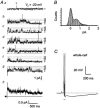
Figure 1. Afterhyperpolarization (AHP) channels in a cell-attached patch recording from an electrophysiologically identified AH neuron
Aa, the pipette potential was held at −60 mV and then stepped to −50 mV. Few channel openings were observed in the patch in the absence of action potential firing: c, closed state; o1, one channel open. b, stepping the pipette potential (Vp) from −60 mV to +60 mV for 110 ms evoked a single action current (arrow) in the neuron and this was followed by the opening of up to three channels at VP = −50 mV. Ba and b, all-points histograms of the corresponding traces shown in A. Note the increase in Po following AP firing. Unitary current amplitude was 0.37. c, i-V relationship of KAHP channels at three values of Vp. A linear regression line through the data points gave a slope conductance of approximately 40 pS. C, whole-cell current clamp recording from the same neuron which generated a slow AHP following a three-pulse stimulus train. The bath contained Krebs solution and the pipette was filled with 145 mm KCl, containing 50 nm Ca2+.
Figure 2
Figure 2. Cell-attached patch recording from an AH neuron with BK and SK channels blocked with TEA (5 mm) and apamin (0.5 μm)
Aa, in the absence of APs, few channel openings were evident (the asterisk marks the opening of one channel). VP = −20 mV. Traces b-g were recorded following 110 ms step depolarizations of the patch (VP = +60 mV) to trigger an AP in the neuron (not shown). Following AP firing, there is an increase in channel activity in the patch. In traces d and e, channel openings continued past the end of the recording. Up to four TEA- and apamin-resistant channels are seen to open simultaneously. h, ensemble current averaged from 10 traces containing channel openings. The patch current during the step depolarization has been removed for clarity. Note slow decay of current. B, all-points histogram constructed from traces containing channel openings, revealing three equidistant peaks and fitted with Gaussian curves (closed state on left and two open states; the third open state is within the baseline). Unitary current was 0.67 pA. C, whole-cell recording after patch rupture shows prominent slow AHP following three evoked action potentials (filled bar) recorded under current clamp.
Figure 3
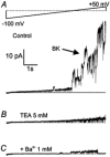
Figure 3. Outside-out patch recording from AH neurons showing two populations of KCa channels
The bath was perfused with Krebs solution and the patch pipette was filled with 145 mm KCl containing 500 nm Ca2+. A, the outside-out patch was ramp-depolarized from −100 mV to +50 mV over 10 s (upper trace). Note activation of BK-type channels (three channels open simultaneously) at potentials positive to approximately +20 mV in the current recording (lower trace). B, ramp current elicited after TEA (5 mm) was added to the Krebs solution. Openings of BK-type channels were abolished, but openings of the smaller conductance channels persisted. C, following the addition of Ba2+ (1 mm) to the TEA-containing Krebs solution, the opening of the smaller conductance channels was markedly inhibited at potentials negative to 0 mV.
Figure 4
Figure 4. Activation by cytoplasmic Ca2+ of TEA- and apamin-resistant KCa channels in an inside-out patch
Aa, two TEA- and apamin-resistant KCa channels were stimulated to open following wash-in of 500 nm Ca2+-containing high-K+ PS. The patch was held at 0 mV under asymmetric K+ concentrations at room temperature (5 mm potassium gluconate in pipette; 145 mm potassium gluconate in bath). The pipette solution contained 5 mm TEA and 0.5 μm apamin. b, time course of activation of KCa channels by Ca2+ expressed as NPo (where N is number of channels), showing persistent activity after wash-out of 500 nm Ca2+ PS. The dotted lines represent gaps in continuous recordings where recordings at other potentials were executed. B, activity of KCa channels before (a) and in the presence of (b) Ca2+ PS. c, activity of KCa channels persisted following 5 min wash-out of Ca2+ PS. d, wash-in of 0 Ca2+ PS containing 145 mm sodium gluconate blocked current through KCa channels.
Figure 5
Figure 5. Rapid activation and deactivation of BK-type channels by Ca2+-PS in another inside-out patch
A, the patch which contained a single BK-type channel was held at 0 mV under asymmetrical transmembrane K+ concentrations. Upon wash-in of Ca2+-containing (10 μm) high-K+ PS, there was a rapid increase in the Po of the BK channel; activity quickly reversed following wash-in of 0 Ca2+ PS. B, recordings from the same patch in response to ramp depolarization in the presence of 0 Ca2+ PS in the bath (a) and after wash-in of 500 nm Ca2+(b). PS ramp depolarization activated BK channels at potentials positive to about +20 mV. c, in the presence of 10 μm Ca2+-containing PS in the bath, the ramp depolarization activated BK channels at potentials positive to about −80 mV. TEA was omitted from the pipette solution.
Figure 6
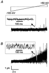
Figure 6. The effect of ramp depolarization of an inside-out patch containing TEA- and apamin-resistant KCa channels
A, ramp depolarization from −100 to +50 mV over 10 s failed to activate channel openings except at very positive potentials (cytoplasmic [Ca2+] < 10 nm). B, following equilibration in 500 nm Ca2+-containing PS on the cytoplasmic side of the patch, ramp depolarization of the patch activated TEA- and apamin-resistant KCa channels throughout the ramp. Insets in each panel show expanded portions of the ramp currents around 0 mV (continuous line indicates closed channel level).
Figure 7
Figure 7. Lack of voltage dependence of opening of TEA- and apamin-resistant KCa channels
A, inside-out patch containing an estimated two channels which were activated with 500 nm Ca2+ high-K+ PS. Steady-state activity is shown at three potentials. The asterisk in trace b indicates the possible simultaneous opening of two channels. o, open state; c, closed state. B, all-points histograms of 20 s recordings of channel activity at the three potentials, fitted with superimposed Gaussian curves. Note that the curves representing the open state distribution are of roughly equal relative area. C, Po-V plot constructed from the mean data, from recordings obtained under asymmetrical K+ concentrations. Data points have been fitted with a Boltzmann function, of the form: Po = Po,max/[1 + exp(V0.5/Vslope)] + Po,basal, where Vslope = 40 mV, V0.5 is −20 mV, Po,max was 0.56 and Po,basal was 0.37. D, i-V relationship of TEA- and apamin-resistant KCa channels under asymmetric K+ concentrations. Data points have been fitted with a form of the GHK current equation, showing open channel outward current rectification. From the fitted curve, the limiting slope conductance at positive potentials is approximately 40 pS and the chord conductance at −50 mV is 12 pS.
Figure 8
Figure 8. Ca2+-dependent activity of TEA- and apamin-resistant KCa channels under symmetrical K+ concentrations
A, wash-in of 5 μm Ca2+-containing PS on an inside-out patch held at −60 mV and containing a single TEA- and apamin-resistant KCa channel, resulted in an increase in channel activity. B, all-points histogram plots of channel activity recorded in 0 Ca2+ PS and in the presence of 5 μm Ca2+, at −60 mV, fitted with superimposed Gaussian curves. Note basal activity of KCa channels (left graph). C, Po-V curve constructed from the mean data collected from three to eight patches exposed to 2–5 μm cytoplasmic Ca2+. Data points have been fitted with a straight line indicating no voltage dependence. D, i-V plot of the mean unitary current ± s.e.m., fitted with a linear regression line between −60 and +60 mV, yielding a unitary conductance of 62 pS.
Figure 9
Figure 9. Kinetic analysis of TEA- and apamin-resistant KCa channel activity recorded under asymmetrical K+ concentrations
Aa, activity in a typical inside-out patch at 0 mV in 0 Ca2+ PS; b, steady-state activity following wash-in of high-Ca2+ PS on the cytoplasmic surface. The trace was digitally filtered at 1 kHz and contains two channels, but simultaneous openings were rare (c). Amplitude histogram plot of the idealized transitions to the open state (o) and to the closed state (c), each fitted with a Gaussian curve. The single channel current was 1.07 and the Po was 0.39. B, dwell time distributions showing that the open dwell time was fitted with a single exponential curve with a time constant of 1.25 ms (a), while the closed dwell time was best fitted with the sum of two exponential functions having time constants of 1.44 and 7.89 ms (b).

Similar articles

Cited by

References

    1. Bekkers JM. Distribution of slow AHP channels on hippocampal CA1 pyramidal neurons. Journal of Neurophysiology. 2000;83:1756–1759. - PubMed
    1. Bertrand PP, Kunze WA A, Bornstein JC, Furness JB. Electrical mapping of the projections of intrinsic primary afferent neurons to the mucosa of the guinea-pig small intestine. Neurogastroenterology. 1998;10:533–541. - PubMed
    1. Bond CT, Maylie J, Adelman JP. Small-conductance calcium-activated potassium channels. Annals of the New York Academy of Sciences. 1999;868:370–378. - PubMed
    1. Cloues RK, Tavalin SJ, Marrion NV. Beta-adrenergic stimulation selectively inhibits long-lasting L-type calcium channel facilitation in hippocampal pyramidal neurons. Journal of Neuroscience. 1997;17:6493–6503. - PMC - PubMed
    1. Cordoba-Rodriguez R, Moore KA, Kao JPY, Weinreich D. Calcium regulation of a slow post-spike hyperpolarization in vagal afferent neurons. Proceedings of the National Academy of Sciences of the USA. 1999;96:7650–7657. - PMC - PubMed

Publication types

LinkOut - more resources