. 2002 Jan 15;538(Pt 2):619-31.
doi: 10.1113/jphysiol.2001.012888.

Sleep-waking discharge patterns of neurons recorded in the rat perifornical lateral hypothalamic area

Affiliations

Sleep-waking discharge patterns of neurons recorded in the rat perifornical lateral hypothalamic area

Md Noor Alam et al. J Physiol. .

Abstract

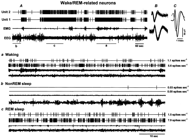
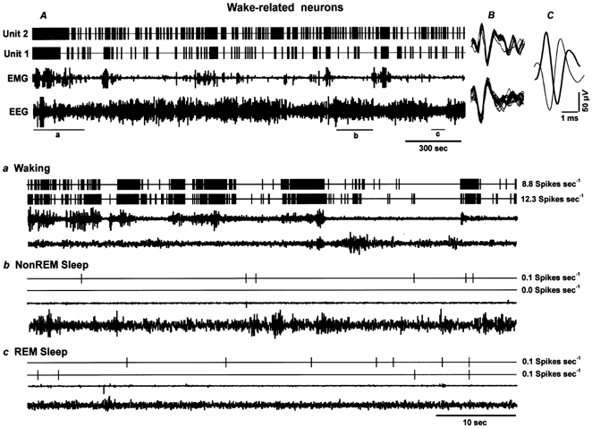
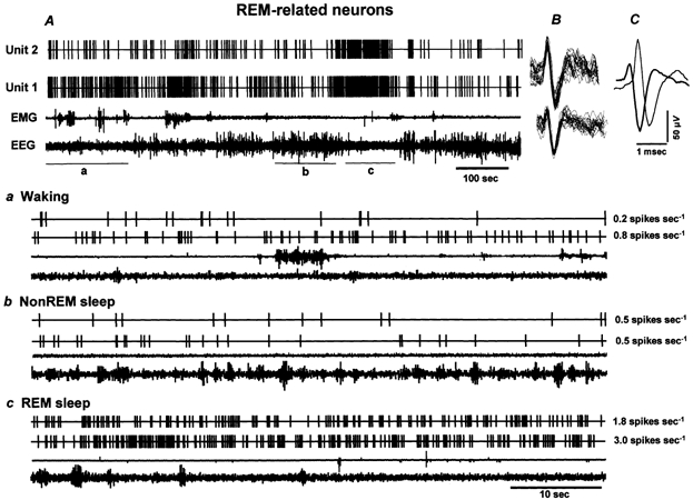
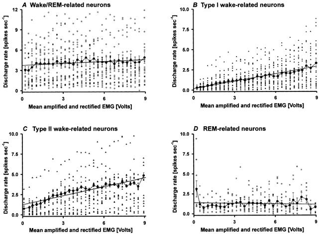
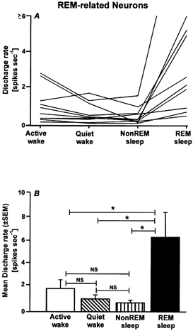
The perifornical lateral hypothalamic area (PF-LHA) has been implicated in the control of several waking behaviours, including feeding, motor activity and arousal. Several cell types are located in the PF-LHA, including projection neurons that contain the hypocretin peptides (also known as orexins). Recent findings suggest that hypocretin neurons are involved in sleep-wake regulation. Loss of hypocretin neurons in the human disorder narcolepsy is associated with excessive somnolence, cataplexy and increased propensity for rapid eye movement (REM) sleep. However, the relationship of PF-LHA neuronal activity to different arousal states is unknown. We recorded neuronal activity in the PF-LHA of rats during natural sleep and waking. Neuronal discharge rates were calculated during active waking (waking accompanied by movement), quiet waking, non-REM sleep and REM sleep. Fifty-six of 106 neurons (53 %) were classified as wake/REM-related. These neurons exhibited peak discharge rates during waking and REM sleep and reduced discharge rates during non-REM sleep. Wake-related neurons (38 %) exhibited reduced discharge rates during both non-REM and REM sleep when compared to that during waking. Wake-related neurons exhibited significantly higher discharge rates during active waking than during quiet waking. The discharge of wake-related neurons was positively correlated with muscle activity across all sleep-waking states. Recording sites were located within the hypocretin-immunoreactive neuronal field of the PF-LHA. Although the neurotransmitter phenotype of recorded cells was not determined, the prevalence of neurons with wake-related discharge patterns is consistent with the hypothesis that the PF-LHA participates in the regulation of arousal, muscle activity and sleep-waking states.


Figures

Figure 1
Figure 1. Distribution of Hcrt-immunoreactive neurons and locations of recorded neurons in the rat PF-LHA
A, photomicrograph of a representative coronal section through the PF-LHA showing the microwire tracts (long arrows) and Hcrt-1-immunoreactive neurons (short arrows). Scale bar, 100 μm. B, camera lucida drawing of a representative coronal section through the PF-LHA showing the distribution of Hcrt-immunoreactive neurons on the right side (•). The area delineated by the thin line indicates the highest density of Hcrt neurons in PF-LHA regions where neuronal recordings were performed. This area has been reproduced on the left side in B and C to indicate the location of recorded neurons with respect to Hcrt neurons. The locations of Type I wake-related (▴) and Type II wake-related (▵) neurons are plotted on the left side in B. Note that nearly all of the recorded wake-related neurons were localized in or immediately adjacent to the Hcrt-immunoreactive neuronal field. C, camera lucida drawing of the same section depicted in B, showing the distribution of wake/REM-related neurons (○) on the left side and REM-related neurons (*) on the right side. Abbreviations: 3V, third ventricle; f, fornix; opt, optic tract.
Figure 2
Figure 2. The discharge pattern of individual wake/REM-related neurons across the sleep-waking cycle
A, 15 min of continuous recording showing the discharge of two wake/REM-related neurons (Units 1 and 2) recorded simultaneously across the sleep-waking cycle. B, the superimposed waveforms of individual action potentials of the recorded neurons captured during five consecutive seconds along with the background signal variations. C, the averaged waveforms of the two action potentials recorded for five consecutive seconds. Note the difference in the spike shape and the duration of the action potentials, even though these neurons were recorded simultaneously from the same microwire. Lower panels illustrate expanded tracings (60 s) from A, showing the discharge patterns of the recorded neurons during waking (a), non-REM sleep (b) and REM sleep (c). The values to the right of each expanded tracing represent mean discharge rates (spikes s−1) for the respective neurons during the representative sections. These neurons exhibited high discharge rates during waking and REM sleep and dramatically reduced discharge rates during non-REM sleep. EEG, electroencephalogram; EMG, neck electromyogram.
Figure 4
Figure 4. Discharge patterns of wake-related neurons across the sleep-waking cycle
A, 66 min tracing showing continuous discharge of two Type I wake-related neurons (Units 1 and 2) recorded simultaneously across the sleep-waking cycle. The superimposed waveforms of individual action potentials captured during 5 s along with the background noise and their averaged waveforms are shown in B and C, respectively. Lower panels depict expanded tracings (60 s) showing discharge rates of these two neurons during waking (a), non-REM sleep (b), and REM sleep (c). Wake-related neurons exhibited highest discharge rates during AW, as indicated by desynchronized EEG and increased EMG activity, a dramatic decline in discharge rates during QW and a further decline during non-REM sleep and REM sleep. The values to the right of each expanded tracing represent the mean discharge rate during the representative sections of the tracing.
Figure 6
Figure 6. The discharge pattern of individual REM-related neurons across the sleep-waking cycle
A, 15 min continuous recording showing the discharge of two REM-related neurons (Units 1 and 2) recorded simultaneously across the sleep-waking cycle. The superimposed waveforms of individual action potentials captured during 5 s for each recorded neuron along with background noise and their averaged waveforms are shown in B and C, respectively. Lower panels depict expanded tracings (60 s) from A, illustrating the discharge patterns of the two neurons during waking (a), non-REM sleep (b) and REM sleep (c). The values to the right of each expanded tracing represent mean discharge rates of the respective neurons during the representative sections. These neurons exhibited lower discharge rates during waking and non-REM sleep and increased discharge rates during REM sleep.
Figure 8
Figure 8. Correlation of PF-LHA neuronal activity with muscle activity
Correlation between discharge rate and EMG activity of wake/REM-related (A), Type I wake-related (B), Type II wake-related (C) and REM-related (D) neurons. Mean (± s.e.m.) discharge rates of individual neurons (large filled circles) are plotted against mean rectified and amplified EMG voltages calculated during successive 5 s epochs across 3–5 sleep-waking cycles. Discharge rates were divided into 0.3 V bins. The straight line in each graph represents the simple linear regression between discharge rate and EMG voltage. The discharge rates of Type I and II wake-related neurons (B and C) were strongly positively correlated with EMG voltage across all sleep-waking states. The discharge rates of a comparable number of wake/REM-related neurons (n = 23) and of REM-related neurons were not significantly correlated with EMG voltage.
Figure 3
Figure 3. Discharge characteristics of wake/REM-related neurons
Individual (A) and mean (± s.e.m.; B) discharge rates (spikes s−1) of wake/REM-related neurons during active waking (AW), quiet waking (QW), non-REM sleep and REM sleep. As a group, these neurons exhibited a significantly higher discharge rate during both AW and REM sleep when compared to QW and non-REM sleep. * P < 0.01. NS, not significantly different.
Figure 5
Figure 5. Discharge characteristics of Type I and II wake-related neurons
Activity of Type I (A and B) and II (C and D) wake-related neurons during AW, QW, non-REM sleep and REM sleep, showing individual (A and C) and mean (± s.e.m.; B and D) discharge rates. Note that both types of neuron exhibited significantly higher discharge rates during AW compared to QW, non-REM and REM sleep. For both types of neuron, discharge rates during QW, non-REM and REM sleep were not significantly different. * P < 0.01.
Figure 7
Figure 7. Discharge characteristics of REM-related neurons
Individual (A) and mean (± s.e.m.; B) discharge rates of REM-related neurons during AW, QW, non-REM sleep and REM sleep. These neurons exhibited low discharge rates during AW, QW and non-REM sleep, with comparatively elevated discharge rates during REM sleep. * P < 0.01.

References

    1. Alam MN, McGinty D, Szymusiak R. Thermosensitive neurons of the diagonal band in rats: relation to wakefulness and non-rapid eye movement sleep. Brain Research. 1997;752:81–89. - PubMed
    1. Aston-Jones G, Bloom FE. Activity of norepinepherine-containing locus coeruleus neurons in behaving rats anticipates fluctuations in the sleep-waking cycle. Journal of Neuroscience. 1981;1:876–886. - PMC - PubMed
    1. Bittencourt JC, Frigo L, Rissman RA, Casatti CA, Nahon JL, Bauer JA. The distribution of melanin-concentrating hormone in the monkey brain (Cebus apella) Brain Research. 1998;804:140–143. - PubMed
    1. Bittencourt JC, Presse F, Arias C, Peto C, Vaughan J, Nahon JL, Vale W, Sawchenko PE. The melanin-concentrating hormone system of the rat brain: an immuno- and hybridization histochemical characterization. Journal of Comparative Neurology. 1992;319:218–245. - PubMed
    1. Bourgin P, Huitron-Resendiz S, Spier AD, Fabre V, Morte B, Criado JR, Sutcliffe JG, Henriksen SJ, De Lecea L. Hypocretin-1 modulates rapid eye movement sleep through activation of locus coeruleus neurons. Journal of Neuroscience. 2000;20:7760–7765. - PMC - PubMed

Publication types

LinkOut - more resources