

Mutational analysis of the interaction between albumin-binding domain from streptococcal protein G and human serum albumin
- PMID: 11790830
- PMCID: PMC2373446
- DOI: 10.1110/ps.02802
Mutational analysis of the interaction between albumin-binding domain from streptococcal protein G and human serum albumin
Abstract
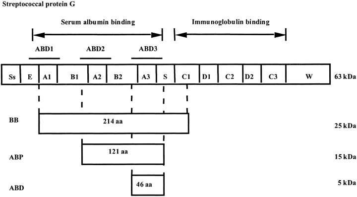
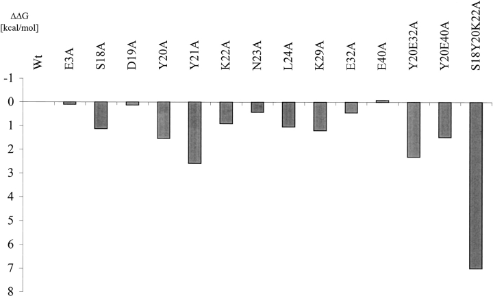
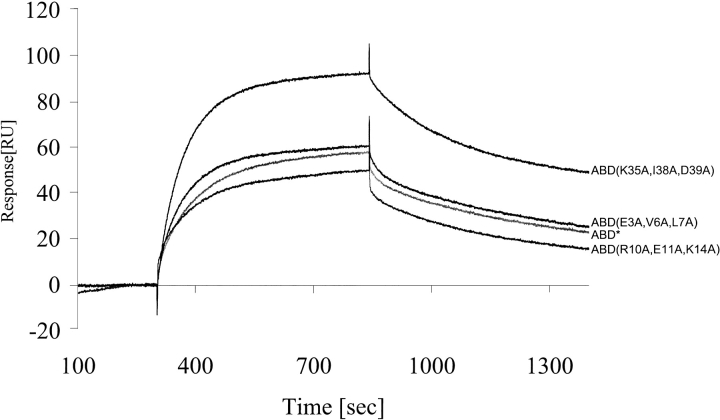
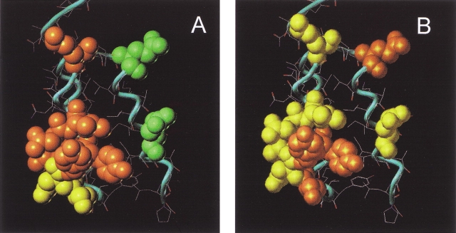
Streptococcal protein G (SpG) is a bacterial cell surface receptor exhibiting affinity to both human immunoglobulin (IgG) and human serum albumin (HSA). Interestingly, the serum albumin and immunoglobulin-binding activities have been shown to reside at functionally and structurally separated receptor domains. The binding domain of the HSA-binding part has been shown to be a 46-residue triple alpha-helical structure, but the binding site to HSA has not yet been determined. Here, we have investigated the precise binding region of this bacterial receptor by protein engineering applying an alanine-scanning procedure followed by binding studies by surface plasmon resonance (SPR). The secondary structure as well as the HSA binding of the resulting albumin-binding domain (ABD) variants were analyzed using circular dichroism (CD) and affinity blotting. The analysis shows that the HSA binding involves residues mainly in the second alpha-helix.
Figures
Similar articles
-
Analysis and use of the serum albumin binding domains of streptococcal protein G.J Mol Recognit. 1988 Apr;1(2):69-74. doi: 10.1002/jmr.300010204. J Mol Recognit. 1988. PMID: 3273653
-
C repeats of the streptococcal M1 protein achieve the human serum albumin binding ability by flanking regions which stabilize the coiled-coil conformation.Biochemistry. 1997 Jul 1;36(26):8107-13. doi: 10.1021/bi962991s. Biochemistry. 1997. PMID: 9201959
-
FcRn binding properties of an abnormal truncated analbuminemic albumin variant.Clin Biochem. 2010 Mar;43(4-5):367-72. doi: 10.1016/j.clinbiochem.2009.12.001. Epub 2009 Dec 16. Clin Biochem. 2010. PMID: 20006594
-
Engineered protein scaffolds for molecular recognition.J Mol Recognit. 2000 Jul-Aug;13(4):167-87. doi: 10.1002/1099-1352(200007/08)13:4<167::AID-JMR502>3.0.CO;2-9. J Mol Recognit. 2000. PMID: 10931555 Review.
-
The albumin-binding domain as a scaffold for protein engineering.Comput Struct Biotechnol J. 2013 Sep 1;6:e201303009. doi: 10.5936/csbj.201303009. eCollection 2013. Comput Struct Biotechnol J. 2013. PMID: 24688717 Free PMC article. Review.
Cited by
-
Identification of B- and T-cell epitopes of BB, a carrier protein derived from the G protein of Streptococcus strain G148.Clin Diagn Lab Immunol. 2003 Jan;10(1):125-32. doi: 10.1128/cdli.10.1.125-132.2003. Clin Diagn Lab Immunol. 2003. PMID: 12522050 Free PMC article.
-
Comparative Preclinical Evaluation of HER2-Targeting ABD-Fused Affibody® Molecules 177Lu-ABY-271 and 177Lu-ABY-027: Impact of DOTA Position on ABD Domain.Pharmaceutics. 2021 Jun 7;13(6):839. doi: 10.3390/pharmaceutics13060839. Pharmaceutics. 2021. PMID: 34200197 Free PMC article.
-
Albumin-binding domain conjugate for near-infrared fluorescence lymphatic imaging.Mol Imaging Biol. 2012 Jun;14(3):301-14. doi: 10.1007/s11307-011-0499-x. Mol Imaging Biol. 2012. PMID: 21688052 Free PMC article.
-
Therapeutic Potential of Bioactive Compounds from Edible Mushrooms to Attenuate SARS-CoV-2 Infection and Some Complications of Coronavirus Disease (COVID-19).J Fungi (Basel). 2023 Aug 31;9(9):897. doi: 10.3390/jof9090897. J Fungi (Basel). 2023. PMID: 37755005 Free PMC article. Review.
-
Biodistribution of a bispecific single-chain diabody and its half-life extended derivatives.J Biol Chem. 2009 Sep 18;284(38):25612-9. doi: 10.1074/jbc.M109.027078. Epub 2009 Jul 23. J Biol Chem. 2009. PMID: 19628871 Free PMC article.
References
-
- Albeck, S. and Schreiber, G. 1999. Biophysical characterization of the interaction of the beta-lactamase TEM-1 with its protein inhibitor BLIP. Biochemistry 38 11–21. - PubMed
-
- Albeck, S., Unger, R., and Schreiber, G. 2000. Evaluation of direct and cooperative contributions towards the strength of buried hydrogen bonds and salt bridges. J. Mol. Biol. 298 503–520. - PubMed
-
- Bradford, M.M. 1976. A rapid and sensitive method for the quantitation of microgram quantities of protein utilizing the principle of protein-dye binding. Anal. Biochem. 72 248–254. - PubMed
-
- Carter, D.C., and He, X.M. 1990. Structure of human serum albumin. Science 249 302–303. - PubMed
MeSH terms
Substances
LinkOut - more resources
Full Text Sources
Other Literature Sources
