Comparative Study
. 2002 Feb;11(2):221-32.
doi: 10.1110/ps.33202.

Rapid structural fluctuations of the free HIV protease flaps in solution: relationship to crystal structures and comparison with predictions of dynamics calculations

Affiliations
Comparative Study

Rapid structural fluctuations of the free HIV protease flaps in solution: relationship to crystal structures and comparison with predictions of dynamics calculations

Darón I Freedberg et al. Protein Sci. 2002 Feb.

Abstract

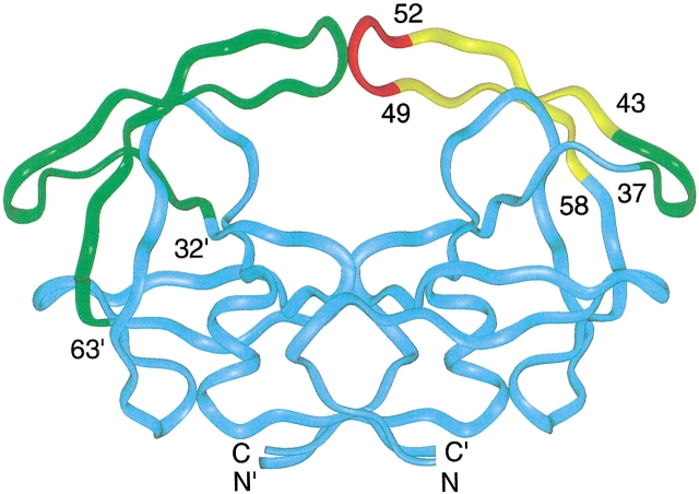
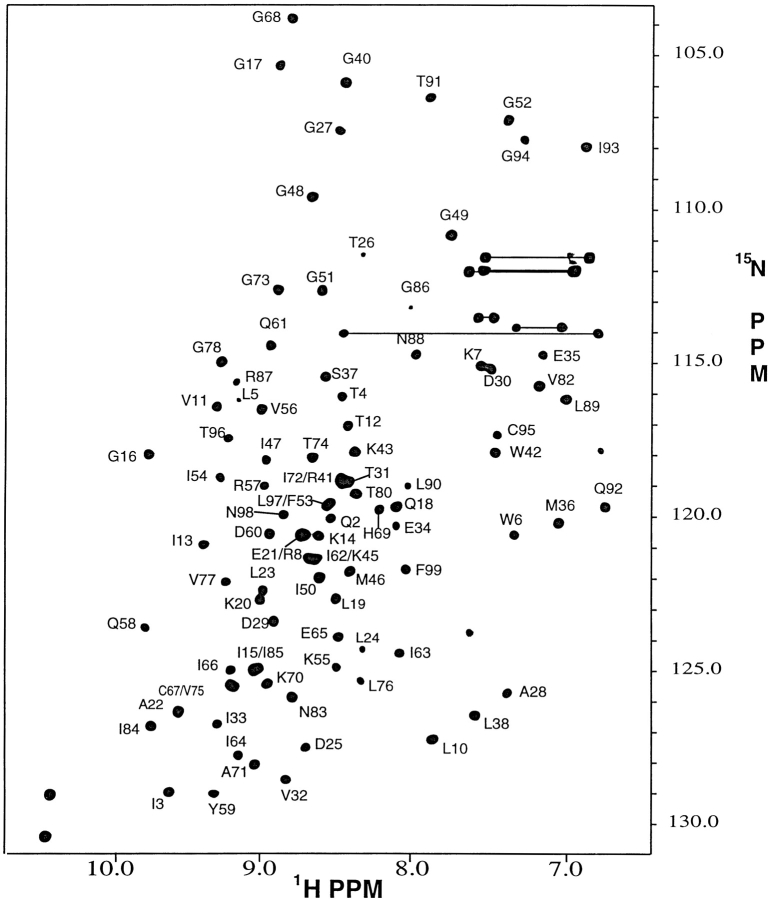
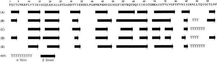
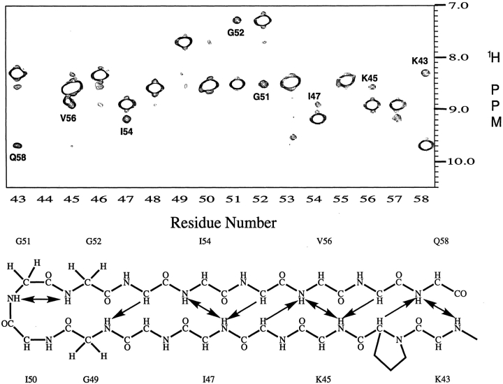
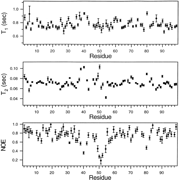
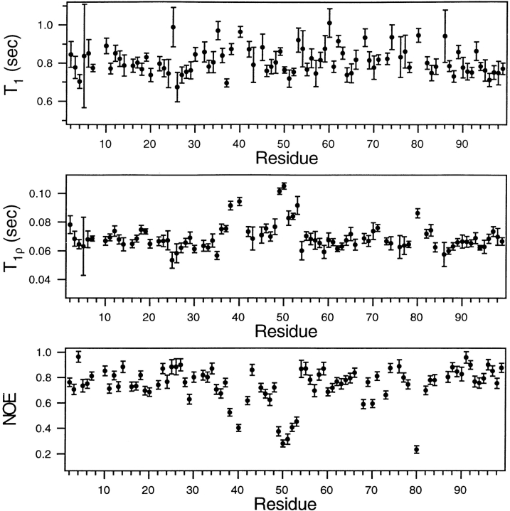
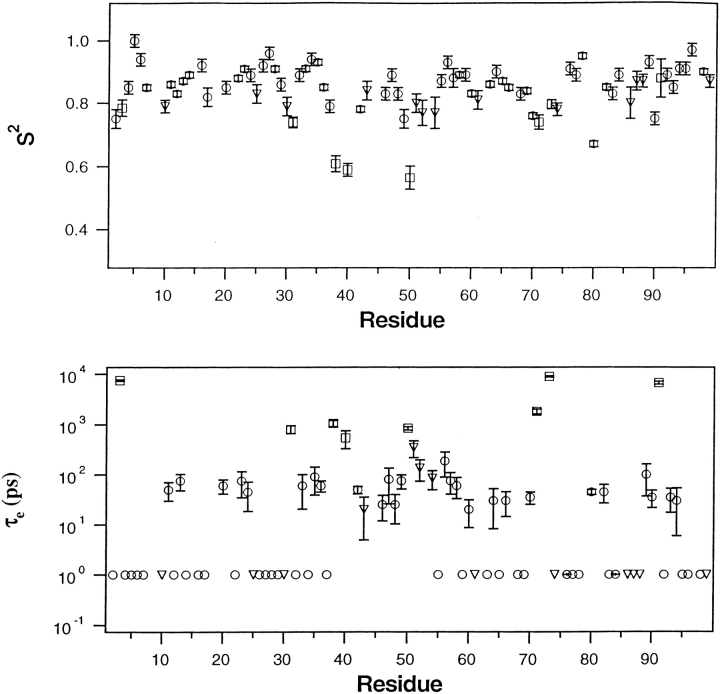
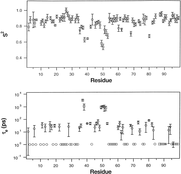
Crystal structures have shown that the HIV-1 protease flaps, domains that control access to the active site, are closed when the active site is occupied by a ligand. Although flap structures ranging from closed to semi-open are observed in the free protease, crystal structures reveal that even the semi-open flaps block access to the active site, indicating that the flaps are mobile in solution. The goals of this paper are to characterize the secondary structure and fast (sub-ns) dynamics of the flaps of the free protease in solution, to relate these results to X-ray structures and to compare them with predictions of dynamics calculations. To this end we have obtained nearly complete backbone and many sidechain signal assignments of a fully active free-protease construct that is stabilized against autoproteolysis by three point mutations. The secondary structure of this protein was characterized using the chemical shift index, measurements of (3h)J(NC') couplings across hydrogen bonds, and NOESY connectivities. Analysis of these measurements indicates that the protease secondary structure becomes irregular near the flap tips, residues 49-53. Model-free analysis of (15)N relaxation parameters, T(1), T(2) (T(1rho)) and (15)N-[(1)H] NOE, shows that residues in the flap tips are flexible on the sub-ns time scale, in contrast with previous observations on the inhibitor-bound protease. These results are compared with theoretical predictions of flap dynamics and the possible biological significance of the sub-ns time scale dynamics of the flap tips is discussed.


Figures

Fig. 1.
Fig. 1.
A colored ribbon representation of the backbone structure of the free HIV-1 protease homodimer. In the left monomer, the flap domain, residues 32`–63` is represented by the green ribbon, whereas the ribbon representing the remaining residues is cyan. In the right monomer, the flap is represented by a tri-colored ribbon: red, residues in the tip of the flap β-hairpin, 49–52; yellow, residues 43–48 and 53–58 in the two-stranded sheet portion of the flap β-hairpin; green, residues 37–42, referred to as the flap elbow. Several residues in the flap domain, are identified by number in the right monomer. The drawing was generated by INSIGHT using the X-ray coordinates PDB accession code 3PHV (Lapatto et al. 1989).
Fig. 2.
Fig. 2.
A 1H– 15N HSQC 500 MHz spectrum of the free HIV-1 protease recorded at pH 5.8 and 25°C. Backbone amide crosspeaks are labeled with the one letter amino acid code and number in the amino acid sequence. Sidechain 15N–1H2 signal pairs of Asn and Gln residues are connected by horizontal lines.
Fig. 3.
Fig. 3.
Comparison of the secondary structure of the free HIV-protease in solution determined by NMR (A) with secondary structures (BE) in four independent crystal structures of the free protein. Secondary structure in solution was identified using the consensus chemical shift index (CSI). The secondary structures in (BE) were derived using PROCHECK (Laskowski et al. 1993) and the following atomic coordinates: (B) Bhat and Erickson, pers. comm.; (C) 3HVP (Wlodawer et al. 1989); (D) 3PHV (Lapatto et al. 1989); and (E) 1HHP (Spinelli et al. 1991). The β-strands found in solution agree with those found in the crystal structures, at least to the extent that β-strand regions in the crystal structures agree among themselves. The Cα CSI identified a short helix spanning residues 87–90, although the consensus CSI did not.
Fig. 4.
Fig. 4.
(A) Strips taken from the amide proton region of the 500 MHz 3D 15N-edited 1H–1H NOESY spectrum of the free protease, recorded with a mixing time of 70 ms. The strips reveal the amide proton NOE's observed for residues 43–58, which are mapped on to the idealized secondary structure of the β-hairpin depicted in (B). In (A), the intense signal in each strip is the (diagonal) amide NH signal of the amino acid numbered at the bottom of the spectrum. The numbered cross-peak in each strip is a long range dNN connectivity between the diagonal amide proton and the amide proton of the residue identified by the number. In (B), the double arrows indicate amide protons that are linked by a pair of observed dNN connectivities. Single arrows identify long range dαN connectivites, linking α- and amide protons, that were observed in the upfield portion of the NOESY spectrum (not shown). A pair of very weak G49–G52 dNN connectivities, just above the noise level, are observed when the NOESY data are plotted at a lower threshold. Of the total of ten inter-strand hydrogen bonds expected in an ideal β-hairpin flap structure, only one, Q58NH–OC`K43, was identified by the hydrogen bond experiment discussed in the text.
Fig. 5.
Fig. 5.
A comparison of backbone amide 15N T1, T2, and NOE values, measured at 25°C and plotted as a function of amino acid residue number of the free HIV-1 protease. The relaxation parameters and uncertainties shown are the averages of two independent data sets. The errors in the T1, T2, and NOE parameters, when averaged over all residues, are 3.2, 2.7, and 8 %, respectively. The T2 values were recorded using a CPMG sequence, with a 1 ms separation between 180° pulses, corresponding to a 500 Hz effective RF field. As described in Materials and Methods, the NOE's are corrected for incomplete recovery of 1H and 15N magnetization.
Fig. 6.
Fig. 6.
A comparison of backbone amide 15N T1, T2(T), and NOE values, measured at 20°C and plotted as a function of amino acid residue number of the free HIV-1 protease. The T2(T) values were recorded using a 2 kHz spin lock. As described in Materials and Methods, the T values are corrected for off-resonance effects and the NOE's are corrected for incomplete recovery of 1H and 15N magnetization.
Fig. 7.
Fig. 7.
Model-free parameters S2 and τe of backbone amides, derived from a model-free analysis of the 25°C relaxation data presented in Figure 5 ▶. After using T1/T2 values and the X-ray coordinates of the protease structure depicted in Figure 1 ▶, PDB accession code 3PHV, to derive the components of the rotational diffusion tensor describing global tumbling of the protease (Tjandra et al. 1996), a model-free analysis was carried out using D/D = 1.27 and τ = 12.1 ns (Table 1). Satisfactory fits to the data (χ2 < 7) were obtained using: the simple model-free (circle), simple model-free plus exchange (triangle), and local diffusion plus extended model-free (square) as discussed in the text. Within the experimental errors shown, the same values of S2 were obtained for residues 49–53 using either coordinates of the closed flap structure (Bhat and Erickson, pers. comm.) or averages of angles obtained from the coordinates of the four free-protease structures discussed in Figure 3 ▶. In lower panel, a symbol appearing at 1 ps without error bars signifies a τe value of <∼100 ps that has a large uncertainty.
Fig. 8.
Fig. 8.
Model-free parameters S2 and τe of backbone amides, derived from a model-free analysis of the 20°C relaxation data presented in Figure 6 ▶. After using T1/T values and the X-ray coordinates of the protease having a semi-open flap structure, PDB accession code 3PHV, to derive the components of the rotational diffusion tensor describing global tumbling of the protease (Tjandra et al. 1996), a model-free analysis was carried out using D/D = 1.32 and τ = 12.8 ns (Table 1). Satisfactory fits to the data (χ2 < 7) were obtained using: the simple model-free (circle), simple model-free plus exchange (triangle), and local diffusion plus extended model-free (square) as discussed in the text. Within the experimental errors shown, the same values of S2 were obtained for residues 49–53 using either coordinates of the closed flap structure (T. N. Bhat and John Erickson, pers. comm.) or averages of angles obtained from the coordinates of the four free-protease structures discussed in Figure 3 ▶. In lower panel, a symbol appearing at 1 ps without error bars signifies a τe value of <∼100 ps that has a large uncertainty.

References

    1. Bruschweiler, R., Liao, X.B., and Wright, P.E. 1995. Long-range motional restrictions in a multidomain zinc-finger protein from anisotropic tumbling. Science 268: 886–889. - PubMed
    1. Clore, G.M., Szabo, A., Bax, A., Kay, L.E., Driscoll, P.C., and Gronenborn, A. 1990. Deviation from the simple two-parameter model-free approach to the interpretation of nitrogen-15 nuclear magnetic relaxation of proteins. J. Am. Chem. Soc. 112: 4989–4991.
    1. Collins, J.R., Burt, S.K., and Erickson, J.W. 1995. Flap opening in HIV-1 protease simulated by `activated' molecular dynamics. Nat. Struct. Biol. 2: 334–338. - PubMed
    1. Cordier, F. and Grzesiek, S. 1999. Direct observation of hydrogen bonds in proteins by interresidue 3hJNC` scalar couplings. J. Am. Chem. Soc. 121: 1601–1602. - PubMed
    1. Cornilescu, G., Ramirez, B.E., Frank, M.K., Clore, G.M., Gronenborn, A.M., and Bax, A. 1999. Correlation between 3hJNC` and hydrogen bond length in proteins. J. Am. Chem. Soc. 121: 6275–6279.

Publication types