. 2002 Feb;11(2):342-9.
doi: 10.1110/ps.48702.

Low levels of asparagine deamidation can have a dramatic effect on aggregation of amyloidogenic peptides: implications for the study of amyloid formation

Affiliations

Low levels of asparagine deamidation can have a dramatic effect on aggregation of amyloidogenic peptides: implications for the study of amyloid formation

Melanie R Nilsson et al. Protein Sci. 2002 Feb.

Abstract

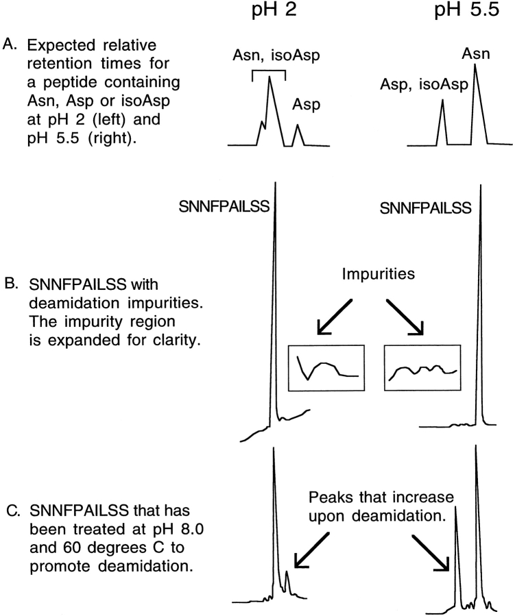
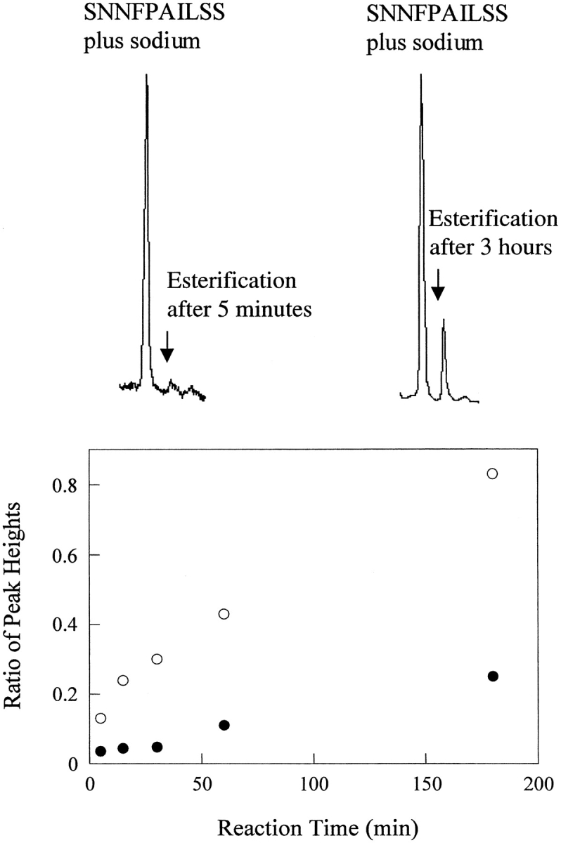
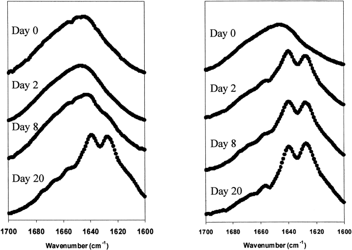
The polypeptide hormone amylin forms amyloid deposits in Type 2 diabetes mellitus and a 10-residue fragment of amylin (amylin(20-29)) is commonly used as a model system to study this process. Studies of amylin(20-29) and several variant peptides revealed that low levels of deamidation can have a significant effect on the secondary structure and aggregation behavior of these molecules. Results obtained with a variant of amylin(20-29), which has the primary sequence SNNFPAILSS, are highlighted. This peptide is particularly interesting from a technical standpoint. In the absence of impurities the peptide does not spontaneously aggregate and is not amyloidogenic. This peptide can spontaneously deamidate, and the presence of less than 5% of deamidation impurities leads to the formation of aggregates that have the hallmarks of amyloid. In addition, small amounts of deamidated material can induce amyloid formation by the purified peptide. These results have fundamental implications for the definition of an amyloidogenic sequence and for the standards of purity of peptides and proteins used for studies of amyloid formation.


Figures

Fig. 1.
Fig. 1.
HPLC traces showing the presence of deamidation impurities. (A) A schematic diagram depicting the relative retention time of Asn, Asp, and iso-Asp containing peptides at pH 2 or pH 5.5. Adapted from Aswad and Guzetta (1995). (B) HPLC traces of the SNNFPAILSS peptide that show the small impurities. The region corresponding to the impurities is expanded in the boxes for clarity. Note that these impurities constitute less than 5% of the total sample. (C) HPLC trace that confirms that the impurities are due to deamidation by demonstrating that the impurity peaks increase upon further deamidation. Additional deamidation was promoted by heating the sample to 60°C at pH 8.0. The prominent deamidation peak at pH 2 is smaller than the peak at pH 5.5. This is because the peak at pH 2 represents only the Asp peptide, while at pH 5.5 in phosphate the peak represents Asp plus iso-Asp [see (A)].
Fig. 2.
Fig. 2.
MALDI-MS detected methyl esterification. The top figures show representative MALDI-MS traces. The major peaks correspond to SNNFPAILSS plus sodium. The minor peaks that result from esterification (after either 5 min or 3 h of reaction with MeOH: HCl) are labeled. The bottom plot shows a time course of methyl esterification of SNNFPAILSS (filled circles) and SNDFPAILSS (open circles) followed by MALDI-MS. The ratio of the 1071 peak to the 1086 peak is plotted as a function of reaction time. The larger change in the ratio for the control SNDFPAILSS sample simply reflects the fact that the entire sample is capable of undergoing esterification. In contrast, only the deamidated material is susceptible to esterification in the SNNFPAILSS peptide.
Fig. 3.
Fig. 3.
FTIR spectra of a sample of SNNFPAILSS that contains no deamidation impurities. (A) FTIR amide I band of SNNFPAILSS recorded at pD 2.29. Experimental data is shown in black circles. Component bands identified by curve fitting are shown as solid lines (1647 cm−1, 1677 cm−1). (B) FTIR amide I band of SNNFPAILSS recorded at pD 5.47. Experimental data is shown in black circles. Component bands identified by curve fitting are shown as solid lines (1647 cm−1, 1677 cm−1). TEM micrographs of these samples contained few ordered deposits and are not shown.
Fig. 4.
Fig. 4.
FTIR spectra and TEM images of a sample of SNNFPAILSS that contains deamidation impurities. (A) FTIR amide I band of SNNFPAILSS recorded in D2O at pD 2.25. Experimental data is shown in black circles. Component bands identified by curve fitting are shown as solid lines (1647 cm−1, 1676 cm−1). TEM micrographs of the pD 2.25 sample showed few ordered deposits and are not shown. (B) FTIR amide I band of SNNFPAILSS recorded at pD 5.71. Experimental data is shown in black circles. Component bands identified by curve fitting are shown as solid lines (1620 cm−1, 1643 cm−1, 1660 cm−1, and 1675 cm−1). (C) TEM micrograph of SNNFPAILSS at pD 5.71 showing amyloid-like fibrils. The scale bar represents 100 nm. (D) TEM micrograph of SNNFPAILSS at pD 5.71 showing the sheet-like assemblies. The scale bar represents 1 μm.
Fig. 5.
Fig. 5.
Time course of seeding experiments followed by FTIR. FTIR spectra of pure SNNFPAILSS (left) and SNNPAILSS seeded with deamidation impurities (right). FTIR spectra were recorded on the day of sample preparation (designated day 0) and on days 1, 2, 3, 4, 8, and 20.

Similar articles

Cited by

References

    1. Ashburn, T.T. and Lansbury, P.T., Jr. 1993. Interspecies sequence variations affect the kinetics and thermodynamics of amyloid formation: Peptide models of pancreatic amyloid. J. Am. Chem. Soc. 11511012–11013.
    1. Ashburn, T.T., Auger, M., 1st., and Lansbury, P.T., Jr. 1992. The structural basis of pancreatic amyloid formation: Isotope-edited spectroscopy in the solid state. J. Am. Chem. Soc. BO114: 790–791.
    1. Aswad, D.W. and Guzetta, A.W. 1995. Methods for analysis of deamidation and isoaspartate formation in peptides and proteins. In Deamidation and isoaspartate formation in peptides and proteins (ed. D.W. Aswad), pp. 8–29. CRC Press, Ann Arbor, MI.
    1. Baumann, M.H., Wisniewski, T., Levy, E., Plant, G.T., and Ghiso, J. 1996. C-terminal fragments of alpha- and beta-tubulin form amyloid fibrils in vitro and associate with amyloid deposits of familial cerebral amyloid angiopathy, British type. Biochem. Biophys. Res. Commun. 219238–242. - PubMed
    1. Betsholtz, C., Christmansson, L., Engstrom, U., Rorsman, F., Jordan, K., O'Brien, T.D., Murtaugh, M., Johnson, K.H., and Westermark, P. 1990. Structure of cat islet polypeptide and identification of amino acid residues of potential significance for islet amyloid formation. Diabetes 39 118–122. - PubMed

Publication types

LinkOut - more resources