. 2002 Jan 22;99(2):996-1001.
doi: 10.1073/pnas.022493799. Epub 2002 Jan 15.

Complete genome sequence of Clostridium perfringens, an anaerobic flesh-eater

Affiliations

Complete genome sequence of Clostridium perfringens, an anaerobic flesh-eater

Tohru Shimizu et al. Proc Natl Acad Sci U S A. .

Abstract

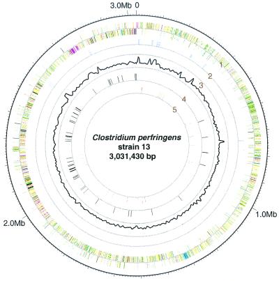
Clostridium perfringens is a Gram-positive anaerobic spore-forming bacterium that causes life-threatening gas gangrene and mild enterotoxaemia in humans, although it colonizes as normal intestinal flora of humans and animals. The organism is known to produce a variety of toxins and enzymes that are responsible for the severe myonecrotic lesions. Here we report the complete 3,031,430-bp sequence of C. perfringens strain 13 that comprises 2,660 protein coding regions and 10 rRNA genes, showing pronounced low overall G + C content (28.6%). The genome contains typical anaerobic fermentation enzymes leading to gas production but no enzymes for the tricarboxylic acid cycle or respiratory chain. Various saccharolytic enzymes were found, but many enzymes for amino acid biosynthesis were lacking in the genome. Twenty genes were newly identified as putative virulence factors of C. perfringens, and we found a total of five hyaluronidase genes that will also contribute to virulence. The genome analysis also proved an efficient method for finding four members of the two-component VirR/VirS regulon that coordinately regulates the pathogenicity of C. perfringens. Clearly, C. perfringens obtains various essential materials from the host by producing several degradative enzymes and toxins, resulting in massive destruction of the host tissues.


Figures

Figure 1
Figure 1
Circular map of the C. perfringens chromosome. The outer circle shows the scale. Ring 1 shows the coding sequence by strand (clockwise and counterclockwise); ring 2 shows the positions of rRNA genes; ring 3 shows G + C content (outer and inner rings correspond to 45% and 20%, respectively); ring 4 shows the positions of sporulation/germination genes; and ring 5 shows the positions of virulence-related genes. We used the following functional categories in ring 1: intermediary metabolism and fermentation (yellow), information pathways (pink), regulatory proteins (sky blue), conserved hypothetical proteins (orange), proteins of unknown function (light blue), insertion sequences and phage-related functions (blue), stable RNAs (purple), cell wall and cell processes (dark green), virulence (red), detoxification and adaptation (white), and sporulation and germination (black).
Figure 2
Figure 2
Relationship between the content of G + A on the leading strand and the percentage of ORFs on the leading strand in various bacteria. The data were calculated from the genomic sequences from B. burgdorferi (Bbu) (38), B. subtilis (Bsu) (26), C. acetobutylicum (Cac) (10),Campylobacter jejuni (Cje) (39), C. perfringens (Cpe), E. coli (Eco) (40), Lactococcus lactis (Lla) (41), M. leprae (Mle) (42), M. tuberculosis (Mtu) (43), Neisseria meningitidis (Nme) (44), Pseudomonas aeruginosa (Pae) (45), Staphylococcus aureus (Sau) (46), Streptococcus pyogenes (Spy) (47), Treponema pallidum (Tpa) (48), and Vibrio cholerae (Vch1) (49). Open and filled circles represent Gram-negative and Gram-positive bacteria, respectively.
Figure 3
Figure 3
Genomic location of the homologous genes in C. perfringens and C. acetobutylicum.
Figure 4
Figure 4
VirR-binding motifs in the C. perfringens genome. The promoter region of the theta-toxin gene is on the top with consensus promoter sequences (−35 and −10) and the mRNA start point (31, 35). The putative VirR-binding sequences (shown in red) are aligned below, and deduced promoter sequences are underlined. The possible consensus VirR-binding motif is shown on the bottom.

References

    1. McDonel J L. Pharmacol Ther. 1980;10:617–635. - PubMed
    1. Hatheway C L. Clin Microbiol Rev. 1990;3:66–98. - PMC - PubMed
    1. Petit L, Gibert M, Popoff M R. Trends Microbiol. 1999;7:104–110. - PubMed
    1. Rood J I. Annu Rev Microbiol. 1998;52:333–360. - PubMed
    1. Rood J I, Cole S T. Microbiol Rev. 1991;55:621–648. - PMC - PubMed

Publication types

MeSH terms