

doi: 10.1073/pnas.022523499.
Epub 2002 Jan 15.
APH-1 is a multipass membrane protein essential for the Notch signaling pathway in Caenorhabditis elegans embryos
Affiliations
- PMID: 11792846
- PMCID: PMC117381
- DOI: 10.1073/pnas.022523499
Item in Clipboard
APH-1 is a multipass membrane protein essential for the Notch signaling pathway in Caenorhabditis elegans embryos
Proc Natl Acad Sci U S A.
.
Abstract
Early embryonic cells in Caenorhabditis elegans embryos interact through a signaling pathway closely related to the Notch signaling pathway in Drosophila and vertebrates. Components of this pathway include a ligand, receptor, the presenilin proteins, and a novel protein, APH-2, that is related to the Nicastrin protein in humans. Here we identify the aph-1 gene as a new component of the Notch pathway in Caenorhabditis elegans. aph-1 is predicted to encode a novel, highly conserved multipass membrane protein. We show that aph-1 and the presenilin genes share a similar function in that they are both required for proper cell-surface localization of APH-2/Nicastrin.
Figures
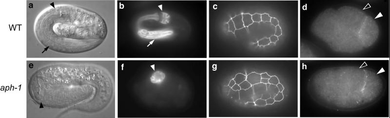
aph-1 and wild-type embryos. (a) Living
wild-type embryo viewed by light microscopy; the arrow indicates the
anterior half of the pharynx and the arrowhead indicates the posterior
half. (b) Immunostaining of pharyngeal cells; markers as
in a. (c) Hypodermal cell boundaries
delineated by green fluorescent protein expression; 10 cells are
visible in the lateral row. (d) Four-cell embryo showing
expression of the ligand APX-1. APX-1 is visible in the posterior-most
cell (P2; white arrowhead) as a line contacting one of the
GLP-1-expressing cells (ABp; black arrowhead); embryos oriented with
the anterior to the left. (e–h)
aph-1 mutant embryos staged, prepared, and labeled as
above. Note the extra hypodermal cells at the first, second, and fourth
positions in g. e and g are
aph-1(zu147), and f and h
are aph-1(zu123).

(a) alignment of the predicted APH-1 protein of
C. elegans with related proteins in
Drosophila (33% identity, GenBank accession no.
AAF51212) and humans (33% and 34% identity, GenBank accession nos.
AAD34072 and AL136671, respectively). Each of the two human genes also
has a highly related mouse gene, which is not shown here (GenBank
accession nos. AC015932.1 and AK002310). Identical amino acids are
black and conserved amino acids are gray; predicted transmembrane
domains are overlined. Asterisks correspond to positions of
aph-1 mutations as follows. zu123, Met to
Ile; or28, Gly to Asp; zu147, Arg to opal
stop codon. (b) APH-1 hydrophobicity plots (21);
alignments as shown in a; predicted membrane-spanning
regions are numbered above the plots.
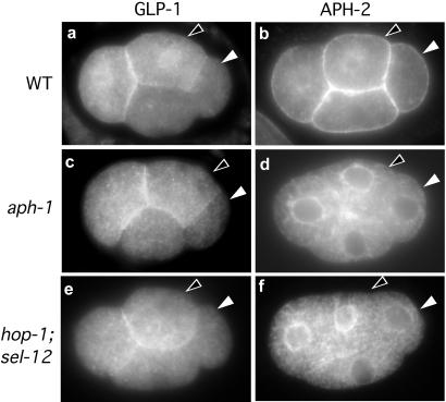
GLP-1 and APH-2 localization in 4-cell stage embryos. The signaling
cell (P2; white arrowhead) and responding cell (ABp; black arrowhead)
are indicated in all panels. (a) Immunostaining of GLP-1
in wild type. GLP-1 is expressed on the surface of the anterior-most
cell (Left) and its sister (black arrowhead).
(b) Immunostaining of APH-2 in wild type. APH-2 is
visible on the surface membranes of all four cells. (c
and d) aph-1(zu123) embryos labeled as
above. (e and f) Embryos
deficient in presenilins [sel-12(ty11);
hop-1(RNAi)] labeled as above. The level of APH-2
immunostaining is highest when cells are mid-interphase; in the images
shown, the GLP-1-expressing cells appear to express a higher level of
APH-2 because they are slightly more advanced in the cell cycle than
the other two cells.
References
-
- Artavanis-Tsakonas S, Rand M D, Lake R J. Science. 1999;284:770–776. - PubMed
-
- Greenwald I. Genes Dev. 1998;12:1751–1762. - PubMed
-
- Kimble J, Simpson P. Annu Rev Cell Dev Biol. 1997;13:333–361. - PubMed
-
- Kopan R, Goate A. Genes Dev. 2000;14:2799–2806. - PubMed
-
- Yu G, Nishimura M, Arawaka S, Levitan D, Zhang L, Tandon A, Song Y Q, Rogaeva E, Chen F, Kawarai T, et al. Nature (London) 2000;407:48–54. - PubMed
Publication types
MeSH terms
Substances
LinkOut - more resources
Full Text Sources
Other Literature Sources
Molecular Biology Databases
