. 2002 Jan 22;99(2):890-5.
doi: 10.1073/pnas.022608799. Epub 2002 Jan 15.

Plasminogen activator inhibitor-1 is a major stress-regulated gene: implications for stress-induced thrombosis in aged individuals

Affiliations

Plasminogen activator inhibitor-1 is a major stress-regulated gene: implications for stress-induced thrombosis in aged individuals

Koji Yamamoto et al. Proc Natl Acad Sci U S A. .

Abstract

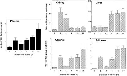
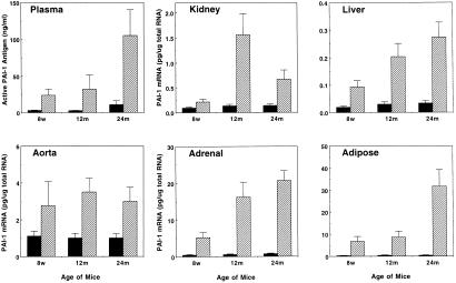
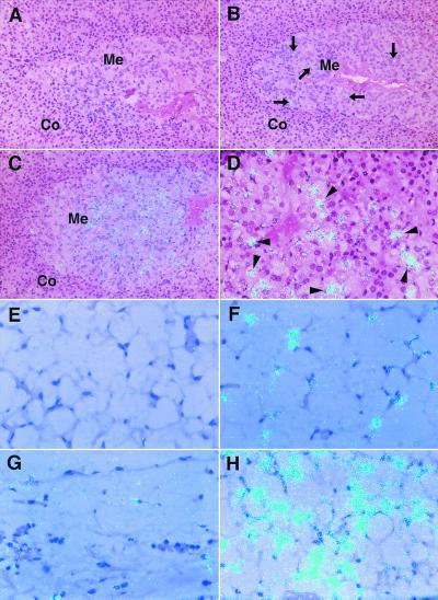
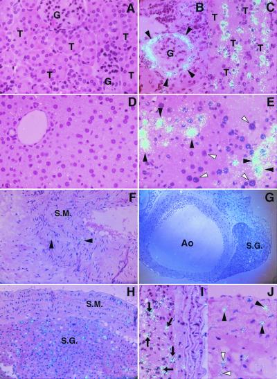
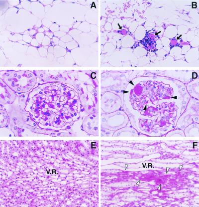
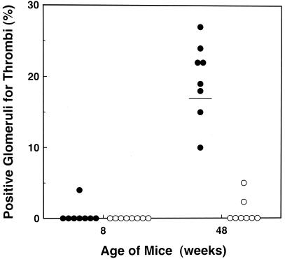
Plasminogen activator inhibitor-1 (PAI-1) is one of the primary inhibitors of the fibrinolytic system and has been implicated in a variety of thrombotic disorders. In this report, stress-induced changes in murine PAI-1 gene expression were investigated to study the role of this inhibitor in the development of stress-induced hypercoagulability. Restraint stress led to a dramatic induction of plasma PAI-1 antigen and of tissue PAI-1 mRNA with maximum induction in adipose tissues. In situ hybridization analysis of the stressed mice revealed that strong signals for PAI-1 mRNA were localized to hepatocytes, renal tubular epithelial cells, adrenomedullar chromaffin cells, neural cells in the paraaortic sympathetic ganglion, vascular smooth muscle cells, and adipocytes, but not to endothelial cells. These observations indicate that the stress induces the PAI-1 gene expression in a tissue-specific and cell type-specific manner. The induction of PAI-1 mRNA by restraint stress was greater than that observed for heat shock protein, a typical stress protein, suggesting that PAI-1 is one of the most highly induced stress proteins. Importantly, the magnitude of induction of PAI-1 mRNA by stress increased markedly with age, and this increase in PAI-1 correlated with tissue thrombosis in the older stressed mice. Moreover, much less tissue thrombosis was induced by restraint stress in young and aged PAI-1-deficient mice compared with age-matched wild-type mice. These results suggest that the large induction of PAI-1 by stress increases the risk for thrombosis in the older populations, and that the adipose tissue may be involved.


Figures

Figure 1
Figure 1
Time course of stress-induced changes in PAI-1 expression in the blood and tissues. Eight-week-old mice were placed into restraint tubes for the indicated times, and then their plasma and tissues were collected. Active PAI-1 antigen levels in the plasma were determined by the tissue plasminogen activator (t-PA) binding assay. Total tissue RNA was prepared and analyzed for PAI-1 mRNA expression level by quantitative RT-PCR. The data are expressed as the mean and SD from six mice in each category.
Figure 2
Figure 2
Comparison of PAI-1 induction by restraint stress between young and aged mice. Plasma and the indicated tissues were collected from 8-week- (8w), 12-month- (12 m), and 24-month (24 m) old mice before (filled bar) and after (hatched bar) 20-h restraint stress. Active PAI-1 antigen levels in the plasma and PAI-1 mRNA expression levels in the tissues were determined by the tissue plasminogen activator (t-PA) binding assay and by quantitative RT-PCR, respectively. The data are expressed as the mean and SD from eight mice in each category.
Figure 3
Figure 3
High resolution in situ hybridization analysis of PAI-1 mRNA in the adrenals and adipose tissues of control and restraint-stressed mice. Adrenals and adipose tissues were harvested from young (8 weeks old) and aged (24 months old) mice before and after 20-h restraint stress, and then analyzed by in situ hybridization by using 35S-labeled cRNA probes as described in Materials and Methods. The hybridization signal for PAI-1 mRNA is indicated by the light blue dots in all panels. (AD) PAI-1 mRNA in adrenals of the unstressed young (A), stressed young (B), and stressed aged mice (C and D). Co, adrenal cortex; Me, adrenal medulla. Arrows in B denote positive cells for PAI-1 mRNA in the adrenal medulla. Arrowheads in D indicate adrenomedullar chromaffin cells. (EH) PAI-1 mRNA in adipose tissues of the unstressed young (E), stressed young (F), unstressed aged (G), and stressed aged mice (H). Magnification: AC, ×200; DH, ×400. All slides were exposed for 10 weeks at 4°C.
Figure 4
Figure 4
High resolution in situ hybridization analysis of PAI-1 mRNA in kidneys, livers, and aortas of control and stressed aged mice. Kidneys, livers, and aortas were harvested from 24-month-old mice before and after 20-h continuous restraint stress, and then analyzed by in situ hybridization as described in Materials and Methods. The hybridization signal for PAI-1 mRNA corresponds to the light blue dots in all panels. (AC) PAI-1 mRNA in the kidneys of the unstressed (A, ×400) and stressed (B and C, ×400) mice. G, glomeruli; T, tubules. Arrowheads indicate epithelial cells in Bowman's capsule (B). (D and E) PAI-1 mRNA in the liver of the unstressed (D, ×400) and stressed (E, ×400) mice. Filled arrowheads denote hepatocytes that strongly expressed PAI-1 mRNA, and open arrowheads indicate negative hepatocytes for PAI-1 mRNA signals (E). (FJ) PAI-1 mRNA in aorta of the unstressed (F) and stressed (GJ) mice. (F) The aortic wall containing smooth muscle layer (S.M.; ×200). (G) The aorta (Ao) and paraaortic sympathetic ganglion (S.G.) at low magnification (×100). (H) The smooth muscle layer (S.M.) of aortic wall and adjacent sympathetic ganglion (S.G.) at higher magnification (×200). (I and J) The aortic wall. Arrows indicate neural cells in the sympathetic ganglion (I, ×400). Filled arrowheads indicate vascular smooth muscle cells, and open arrowheads denote adipocytes (J, ×400). All slides were exposed for 10 weeks at 4°C.
Figure 5
Figure 5
Microscopic analysis on tissue thrombosis induced by restraint stress. Renal and adipose tissues were removed from young (8-week-old) and aged (24-month-old) mice after 20-h restraint stress. Tissue sections were stained with hematoxylin/eosin (A and B), or with PAS (CF), and examined microscopically (n = 6 for each group). (A and B) Adipose tissues of the stressed young (A) and aged (B) mice. Arrows denote thrombi in the microvessels between adipocytes. (CF) Renal tissues of the stressed young (C and E) and aged (D and F) mice. (C and D) A glomerulus. (E and F) The region of vasa recta in the medulla. Filled arrowheads indicate thrombi formation in the glomerulus (D), and open arrowheads denote thrombi in the vasa recta (F). Magnification: A and B, ×200; C and D, ×1,000; E and F, ×400.
Figure 6
Figure 6
Renal glomerular thrombosis in PAI-1-deficient mice and wild-type after 20-h restraint stress. Mice were placed into restraint tubes for 20 h and then killed. The kidneys were removed and analyzed for glomerular thrombosis with hematoxylin/eosin or PAS staining. Stress-induced renal glomerular thrombosis was evaluated quantitatively in young (8 weeks old) wild-type (●) and PAI-1-deficient (○) mice (n = 8), or in older (48 weeks old) wild-type (●) and PAI-1 deficient (○) mice (n = 8). Quantitation was achieved by counting the positive glomeruli for thrombi out of at least 100 glomeruli in each kidney section. The figure shows the percentage of positive glomeruli for thrombi in each mouse. The horizontal bar represents the mean.

References

    1. Selye H. J Clin Endocrinol Metab. 1946;6:117–230. - PubMed
    1. Anathan J, Goldberg A L, Voellmy R. Science. 1986;232:252–254. - PubMed
    1. Minowada G, Welch W J. J Clin Invest. 1995;95:3–12. - PMC - PubMed
    1. Morimoto R I, Sarge K D, Abravaya K. J Biol Chem. 1992;267:21987–21990. - PubMed
    1. Georgopoulos C, Welch W J. Annu Rev Cell Biol. 1993;9:601–634. - PubMed

Publication types

MeSH terms