. 2002 Jan 22;99(2):727-32.
doi: 10.1073/pnas.022628699. Epub 2002 Jan 15.

Requirement of an intact microtubule cytoskeleton for aggregation and inclusion body formation by a mutant huntingtin fragment

Affiliations

Requirement of an intact microtubule cytoskeleton for aggregation and inclusion body formation by a mutant huntingtin fragment

Paul J Muchowski et al. Proc Natl Acad Sci U S A. .

Abstract

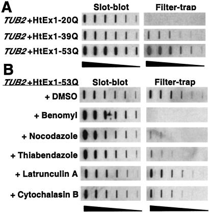
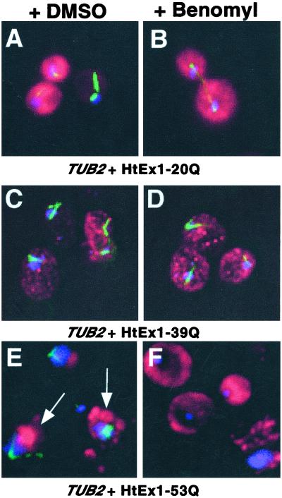
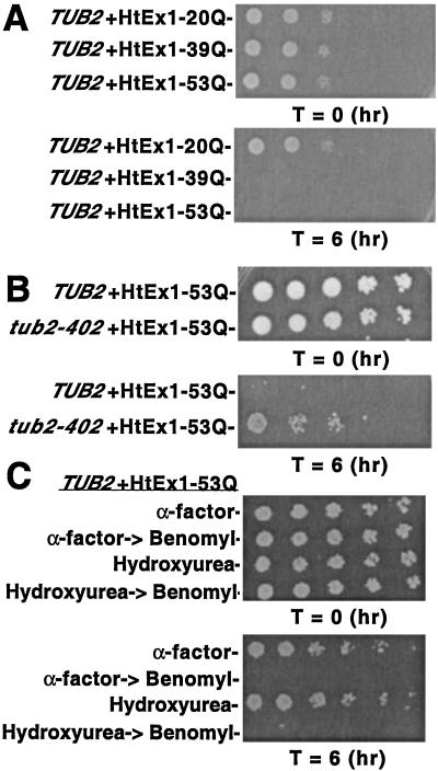
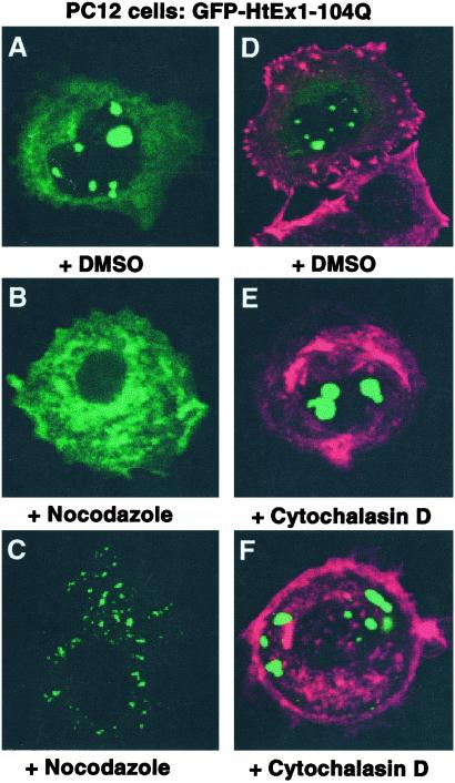
Huntington's disease is caused by the expansion of CAG repeats coding for a polyglutamine tract in the huntingtin protein. The major pathological feature found in Huntington's disease neurons is the presence of detergent-insoluble ubiquitinated inclusion bodies composed of the huntingtin protein. However, the mechanisms that underlie inclusion body formation, and the precise relationship between inclusion bodies and events that initiate toxicity, remain unclear. Here, we analyzed the effects of drugs or genetic mutations that disrupt the microtubule cytoskeleton in a Saccharomyces cerevisiae model of the aggregation of an amino-terminal polyglutamine-containing fragment of huntingtin exon 1 (HtEx1). Treatment of yeast with drugs that disrupt microtubules resulted in less than 2% of the detergent-insoluble HtEx1 observed in mock-treated cells and prevented the formation of large juxtanuclear inclusion bodies. Disruption of microtubules also unmasked a potent glutamine length-dependent toxicity of HtEx1 under conditions where HtEx1 exists in an entirely detergent-soluble nonaggregated form. Results from the yeast model paralleled those from neuronal pheochromocytoma cells, where disruption of microtubules eliminated the formation of juxtanuclear and intranuclear inclusion bodies by HtEx1. Our results suggest that active transport along microtubules may be required for inclusion body formation by HtEx1 and that inclusion body formation may have evolved as a cellular mechanism to promote the sequestration or clearance of soluble species of HtEx1 that are otherwise toxic to cells.


Figures

Figure 1
Figure 1
Requirement of an intact microtubule cytoskeleton for the formation of SDS-insoluble HtEx1-53Q aggregates in yeast. (A) Glutamine length-dependent aggregation of HtEx1 in yeast. Yeast cells (TUB2) were grown and induced for HtEx1 expression with CuSO4 as described in Materials and Methods. After 6 h of induction at 30°C, protein extracts were made and analyzed for HtEx1 expression in slot-blots and for SDS-insoluble aggregates in filter-trap assays. (B) Treatment of yeast with drugs that disrupt microtubules decreases the amount of SDS-insoluble HtEx1-53Q aggregates detected in filter-trap assays. Cells transformed with HtEx1-53Q were grown and treated transiently with microtubule (benomyl, nocodazole, thiabendazole) or actin (latrunculin A, cytochalasin B) drugs as described in Materials and Methods. After 6 h of recovery in media lacking drugs, protein lysates were made and analyzed by slot-blot and filter-trap assays as in A. Shown are two-fold serial dilutions starting with 10 μg of total lysate.
Figure 2
Figure 2
An intact microtubule cytoskeleton is required for the juxtanuclear localization of HtEx1-53Q inclusion bodies in yeast. Cells treated transiently with DMSO (A, C, and E) or benomyl (B, D, and F) were harvested, fixed, and reacted with anti-c-myc and antitubulin Abs to visualize HtEx1 (red) and microtubules (green), respectively, and 4,6-diamino-2-phenylindole (DAPI) to show DNA (blue). (A and B) TUB2 + HtEx1-20Q, (C and D) TUB2 + HtEx1-39Q, (E and F) TUB2 + HtEx1-53Q. The arrows shown in E denote the juxtanuclear localization of HtEx1-53Q.
Figure 3
Figure 3
Transient depolymerization of microtubules with benomyl results in a glutamine length-dependent cell cycle-independent toxicity of HtEx1 in yeast. (A and B) Yeast cells (TUB2 or tub2-402) were treated transiently with 20 μg/ml of benomyl to depolymerize microtubules as described in Materials and Methods. (C) Yeast cells (TUB2) were grown and treated with 5 μM α-factor or 0.1 M hydroxyurea to arrest cells at G1 and S phases of the cell cycle, respectively. After 3 h the cells were harvested, washed with sterile water, and resuspended in fresh media with and without 20 μg/ml of benomyl. Cells without benomyl treatment were allowed to recover for 6 h and then were tested for viability in spotting assays. After 3 h, benomyl-treated cells were harvested, washed in sterile water, resuspended in fresh media, and allowed to recover for 6 h. (AC) Shown are 5-fold serial dilutions starting with equal numbers of cells before (T = 0) and after (T = 6) drug treatment/CuSO4 induction. Cells were spotted on plates containing synthetic complete media ± 400 μM CuSO4 and were incubated at 30°C for 3 days.
Figure 4
Figure 4
Temperature-sensitive yeast strains with mutations of TUB4 (encoding γ-tubulin) accumulate lower levels of SDS-insoluble HtEx1-53Q. Yeast cells (TUB4, tub4-401, or tub4-32) that express HtEx1-53Q were grown in synthetic complete media at 23°C to mid-log phase (OD600 0.5) at which point 400 μM CuSO4 was added to the media to induce HtEx1-53Q expression. After 6 h of incubation at 23°C (permissive temperature, PT) or 37°C (restrictive temperature, RT), protein extracts were prepared by glass bead lysis and analyzed for HtEx1-53Q expression in slot-blots and for SDS-insoluble aggregates in filter-trap assays as in Fig. 1A. Shown are 2-fold serial dilutions starting with 10 μg of total lysate for tub4-1 and parental control strain TUB4 (A) and tub4-32 and parental control strain TUB4 (B).
Figure 5
Figure 5
An intact microtubule cytoskeleton is required for the juxtanuclear and intranuclear localization of GFP-HtEx1-104Q inclusion bodies in PC12 cells. PC12 cells were transfected with pCDNA3-1-GFP-HtEx1-104Q that expresses HtEx1 with 104 glutamine repeats fused to GFP under the control of a cytomegalovirus-based promoter (18). Six hours after transfection, cells were treated with DMSO (A and D), nocodazole (B and C), or cytochalasin D (E and F). After 18 h of incubation at 37°C, cells were fixed and analyzed for GFP (green) or actin (red) fluorescence. B and C and E and F represent two independent examples of PC12 cells that express GFP-HtEx1-104Q and were treated with nocodazole or cytochalasin D, respectively.

References

    1. Zoghbi H Y, Orr H T. Annu Rev Neurosci. 2000;23:217–247. - PubMed
    1. Saudou F, Finkbeiner S, Devys D, Greenberg M E. Cell. 1998;95:55–66. - PubMed
    1. Huynh D P, Figueroa K, Hoang N, Pulst S M. Nat Genet. 2000;26:44–50. - PubMed
    1. Warrick J M, Chan H Y, Gray-Board G L, Chai Y, Paulson H L, Bonini N M. Nat Genet. 1999;23:425–428. - PubMed
    1. Kazemi-Esfarjani P, Benzer S. Science. 2000;287:1837–1840. - PubMed

Publication types

MeSH terms

LinkOut - more resources