Comparative Study
. 2001 Oct;6(4):377-85.
doi: 10.1379/1466-1268(2001)006<0377:goapao>2.0.co;2.

Genomic organization and promoter analysis of the human heat shock factor 2 gene

Affiliations
Comparative Study

Genomic organization and promoter analysis of the human heat shock factor 2 gene

P Nykänen et al. Cell Stress Chaperones. 2001 Oct.

Abstract

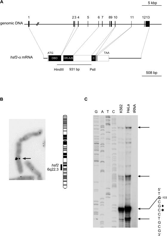
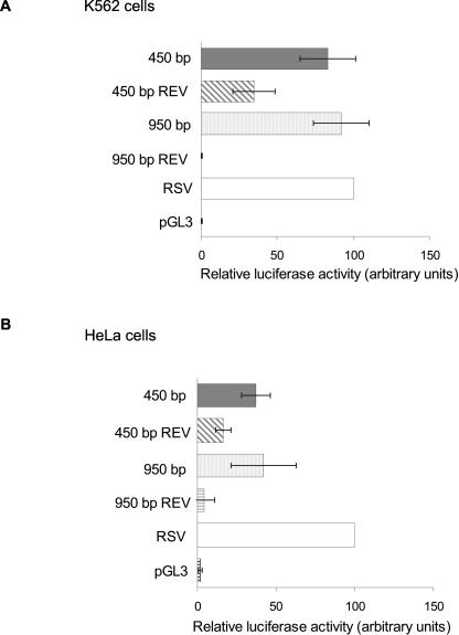
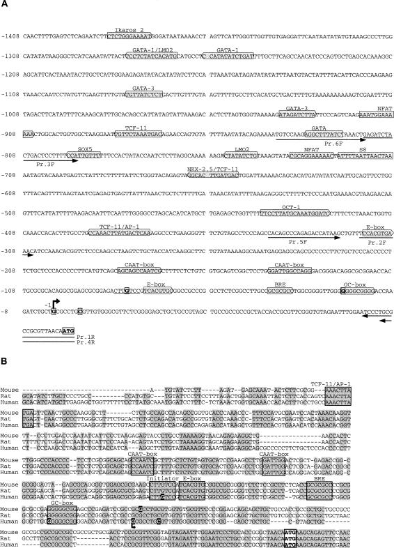
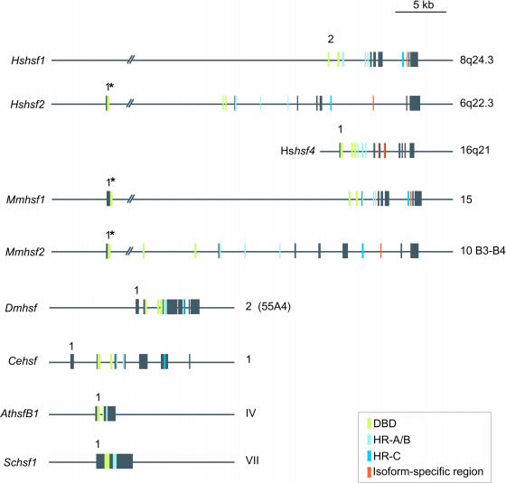
Heat shock factor 2 (HSF2) is a member of the heat shock transcription factor family, which appears to be activated during differentiation and development rather than on cellular stress. Here we report the isolation and characterization of the human hsf2 gene and its 5'-flanking region. The transcription unit of the human hsf2 gene consists of 13 exons dispersed over 33 kbp of genomic DNA on chromosome 6. The hsf2 mRNA is transcribed from multiple start sites, and initiation from the major site results in a transcript of 2.45 kb. A functional promoter, as determined by the ability to direct expression of a transiently transfected luciferase reporter gene, resides in a 950-bp upstream region of the human hsf2 gene. Examination of the core promoter sequence revealed a high GC content and lack of a canonical TATA box. This feature seems to be common among various species, as comparison of the hsf2 proximal promoter sequences from human, mouse, and rat showed distinct conserved regions. Moreover, the overall architecture of the human hsf2 gene is similar to its mouse counterpart. A comparison between human hsf2 gene and other hsf genes showed striking similarities in exon size. However, the exons are assembled in an hsf-specific manner.


Figures

Fig 1.
Fig 1.
The human hsf2 gene. (A) The hsf2 gene spans approximately 33 kbp in the human genome, and the first intron is over 12 kbp. The exons of hsf2 are represented by numbered black boxes and the introns by lines. The functional domains of the HSF2 protein originate from defined groups of exons (DBD: DNA-binding domain; HR-A/B: hydrophobic repeat A/B; HR-C: hydrophobic repeat C). Exon 11 (gray box) is alternatively spliced. The 5′- and 3′-UTRs are 100–200 b and 740 b, respectively. The 931-bp cDNA fragment (Schuetz et al 1991) used as a probe for the P1 screening is shown in the lower panel. The scale of the genomic DNA and mRNA are indicated. (B) To determine the chromosomal localization of the human hsf2 gene, P1 genomic clone was hybridized using fluorescence in situ hybridization to metaphase chromosomes derived from a lymphocyte cell culture. The arrow shows the specific signal of the labeled probe hybridized to chromosome 6, which was identified based on DAPI banding pattern. A schematic representation of human chromosome 6 with the localization of hsf2 is shown. (C) A radiolabeled oligonucleotide, corresponding to positions 13–35 bp upstream of the translation initiation codon, was annealed to total RNA from K562 or HeLa cells or to yeast tRNA. Reverse transcription was carried out, and the extension products were resolved by electrophoresis on a 6% denaturing polyacrylamide gel. A sequencing ladder of human hsf2 5′-flanking region was prepared using the same primer. Arrows indicate the extension products of the most intensive bands obtained in 3 independent experiments. The sequence of the most intense extension product is shown with the start nucleotide in bold (G −103). The asterisk indicates the longest 5′RACE reaction product sequenced, and the diamond indicates the guanine corresponding to the start site deduced for mouse hsf2 (Manuel et al 1999)
Fig 2.
Fig 2.
Luciferase reporter gene analysis of the 5′-flanking region human hsf2. (A) K562 and (B) HeLa cells were transiently transfected with plasmids, in which either a 950-bp or a 450-bp fragment of the 5′-region of the human hsf2 gene was inserted upstream of the luciferase reporter gene in pGL3. The SV40 promoter driving Renilla luciferase gene (SV40-pRL) was transfected into both cell lines as an internal control. Negative controls were provided by vectors containing the promoter fragments in reverse orientation (450 bp REV and 950 bp REV) and by an empty vector (pGL3). The RSV promoter driving the luciferase gene was used as a positive control. The result obtained with RSV was arbitrarily set to 100. The data represent the mean values (±standard deviation) of at least 3 independent experiments in duplicate. All the results are relative to the internal SV40-pRL control plasmid
Fig 3.
Fig 3.
Computer-aided analysis of the 5′-flanking region of the human hsf2 gene. (A) The nucleotide sequence of 1.4 kbp of the human hsf2 5′-flanking region. The ATG codon and the transcription initiation sites are boxed. The first nucleotide upstream of the major transcription initiation site is designated as −1 and indicated with an arrow. Putative transcription factor binding sites, as determined using the MatInspector software V2.2 (Quandt et al 1995) connected to the TRANSFAC database (Heinemeyer et al 1998), are marked by gray boxes, pointing either to the right for binding to the sense strand or to the left for binding to the antisense strand. The quality rating used for choosing the putative transcription factor binding sites is a core similarity of 1.000 and a matrix similarity of ≥0.950. Pr. 1–6 indicate forward (F) and reverse (R) oligonucleotides used for luciferase constructs. (B) Alignment of the proximal promoters of mouse, rat, and human hsf2 promoters by Clustal W 1.8 (−529 bp, −564 bp, and −571 bp relative to ATG, respectively; Thompson et al 1994). The initial methionine ATG codon is shown in the white box, and the identical nucleotides between the different promoters are shaded gray. The 4 transcription initiation nucleotides in human (Fig 1C) and the one determined for mouse (Manuel et al 1999) are highlighted in black background. Arrow marks the major transcription initiation site for human hsf2 (G −103). Data for the transcription initiation sites for rat hsf2 are not available. Putative transcription factor binding sites in the conserved areas are framed
Fig 4.
Fig 4.
Schematic representation of hsf genes. Structures of different hsf genes, as deduced either by comparison between previously published cDNA sequences and genomic sequences available from the GenBank (Hshsf1, Hshsf2, Dmhsf, and Schsf1, http://www.ncbi.nlm.nih.gov) or from previously published genomic structures (Hshsf4, Mmhsf1, and Mmhsf2). The exons are represented by boxes and the introns by lines. Exon 1 is indicated in all genes except in human hsf1. Experimentally determined 5′UTRs are indicated with an asterisk. The chromosomal localization indicated to the right is either verified by experimental data or obtained from various genome projects. The genomic structure of the C. elegans is derived from a computer-generated cDNA sequence compared to the corresponding unspliced genomic sequence found at http://wormbase.sanger.ac.uk. The exons corresponding to the functional domains in the putative C. elegans hsf gene are concluded according to homology to the functional domains of Drosophila. Accession numbers: Hshsf1, M64673 Rabindran et al 1991 (cDNA), AF205589 (genomic); Hshsf2, NM_004506 Schuetz et al 1991 (cDNA), Z99129 (genomic); Hshsf4, NM_001538 Nakai et al 1997 (cDNA), Tanabe et al 1999, AC074143 (genomic); Mmhsf1, X61753 Sarge et al 1991 (cDNA), AF059275/AF061503 Zhang et al 1998 (genomic); Mmhsf2, NM_008297 Sarge et al 1991 (cDNA), AF045615–27 Manuel et al 1999 (exons); Dmhsf, M60070 Clos et al 1990 (cDNA), AE003800 Adams et al 2000 (genomic); Cehsf, AL033536/Y53C10A.12, http://wormbase.sanger.ac.uk (cDNA, genomic); AthsfB1 (Hsf4), Y14069 Prändl et al 1998 (cDNA), Nover et al 2001, Z99707 (genomic) CAB16764 (protein); Schsf-1, M22040 Wiederrecht et al 1988 (cDNA), NC_001139 Tettelin et al 1997 (genomic)

References

    1. Adams MD, Celniker SE, and Holt RA. et al. 2000 The genome sequence of. Drosophila melanogaster. Science. 287:2185–2195. - PubMed
    1. Amin J, Ananthan J, Voellmy R. Key features of the heat shock gene regulatory elements. Mol Cell Biol. 1988;8:3761–3769. - PMC - PubMed
    1. Carey M, Smale ST 2000 Transcription initiation site mapping. In: Transcriptional Regulation in Eukaryotes. Cold Spring Harbor Laboratory Press, Cold Spring Harbor, NY, 116–123.
    1. Clos J, Westwood T, Becker PB, Wilson S, Lambert K, Wu C. Molecular cloning and expression of hexameric Drosophila heat shock factor subject to negative regulation. Cell. 1990;63:1085–1097. - PubMed
    1. Eriksson M, Jokinen E, Sistonen L, Leppä S. Heat shock factor 2 is activated during mouse heart development. Int J Dev Biol. 2000;44:471–477. - PubMed

Publication types

MeSH terms

LinkOut - more resources