

Painful shoulder: comparison of physical examination and ultrasonographic findings
- PMID: 11796399
- PMCID: PMC1754006
- DOI: 10.1136/ard.61.2.132
Painful shoulder: comparison of physical examination and ultrasonographic findings
Abstract
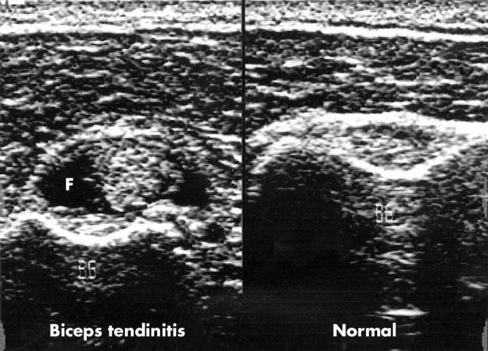
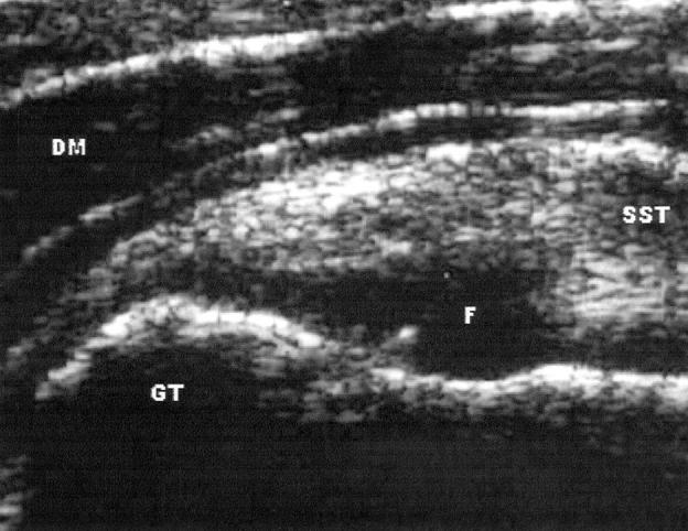
Background: High frequency ultrasonography is an accurate non-invasive imaging technique for evaluating patients with painful shoulder.
Objective: To compare the clinical diagnosis established by a physical examination with high frequency ultrasonographic findings in patients with painful shoulder.
Methods: Thirty one consecutive patients with a first flare of shoulder pain were prospectively included in the study. All had a physical examination performed by two blinded rheumatologists. Ultrasonographic examination was carried out within one week of the physical examination by a third rheumatologist experienced in this technique who had no knowledge of the clinical findings. Ultrasonography was considered the optimal diagnostic technique.
Results: Clinical assessment showed low accuracy in the diagnosis of periarticular shoulder lesions.
Conclusion: Ultrasonography should be used wherever possible to improve diagnosis and treatment of painful shoulder.
Figures
References
Publication types
MeSH terms
LinkOut - more resources
Full Text Sources
Medical
