. 2002 Feb;70(2):434-43.
doi: 10.1128/IAI.70.2.434-443.2002.

CD8(+)-T-cell immunity against Toxoplasma gondii can be induced but not maintained in mice lacking conventional CD4(+) T cells

Affiliations

CD8(+)-T-cell immunity against Toxoplasma gondii can be induced but not maintained in mice lacking conventional CD4(+) T cells

Lori Casciotti et al. Infect Immun. 2002 Feb.

Abstract

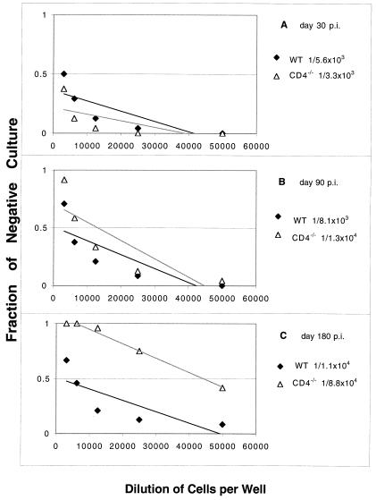
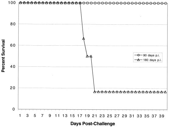
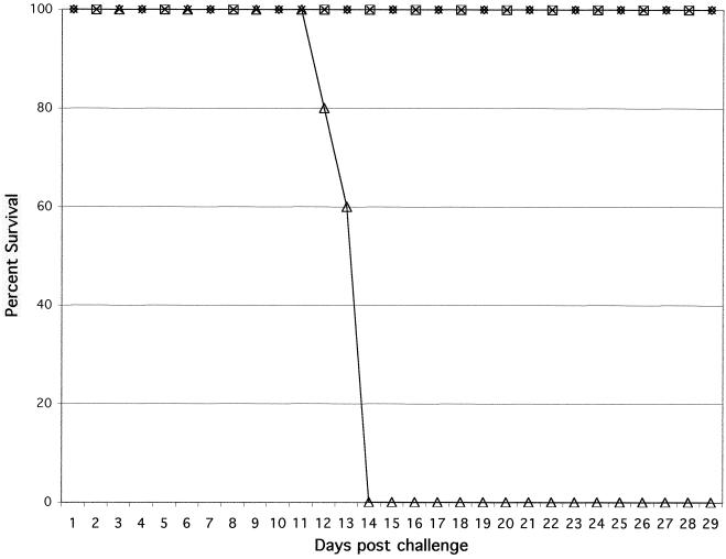
T-cell immunity is critical for survival of hosts infected with Toxoplasma gondii. Among the cells in the T-cell population, CD8(+) T cells are considered the major effector cells against this parasite. It is believed that CD4(+) T cells may be crucial for induction of the CD8(+)-T-cell response against T. gondii. In the present study, CD4(-/-) mice were used to evaluate the role of conventional CD4(+) T cells in the immune response against T. gondii infection. CD4(-/-) mice infected with T. gondii exhibited lower gamma interferon (IFN-gamma) messages in the majority of their tissues. As a result, mortality due to a hyperinflammatory response was prevented in these animals. Interestingly, T. gondii infection induced a normal antigen-specific CD8(+)-T-cell immune response in CD4(-/-) mice. No difference in generation of precursor cytotoxic T lymphocytes (pCTL) or in IFN-gamma production by the CD8(+)-T-cell populations from the knockout and wild-type animals was observed. However, the mutant mice were not able to sustain CD8(+)-T-cell immunity. At 180 days after infection, the CD8(+)-T-cell response in the knockout mice was depressed, as determined by pCTL and IFN-gamma assays. Loss of CD8(+)-T-cell immunity at this time was confirmed by adoptive transfer experiments. Purified CD8(+) T cells from CD4(-/-) donors that had been immunized 180 days earlier failed to protect the recipient mice against a lethal infection. Our study demonstrated that although CD8(+)-T-cell immunity can be induced in the absence of conventional CD4(+) T cells, it cannot be maintained without such cells.


Figures

FIG. 1.
FIG. 1.
Survival of CD4−/− (A) and wild-type (B) mice infected with different doses of T. gondii. Female CD4−/− mice and parental C57BL/6 mice that were 5 to 6 weeks old were challenged perorally with 20, 50, or 100 cysts of T. gondii 76K. Survival of animals was monitored daily. There were six animals per group, and the experiment was performed twice with similar results. The data shown are the data from one of the two experiments.
FIG. 2.
FIG. 2.
IFN-γ mRNA expression following T. gondii infection in CD4−/− and parental wild-type (WT) mice. Mice were infected orally with 20 cysts of T. gondii as described in the text. On day 7 p.i., tissues from both infected (Inf.) and uninfected (Uninf.) controls (three mice per group) were harvested and pooled. Expression of mRNA for IFN-γ was assayed by reverse transcriptase PCR. The transcriptional levels for the genes are expressed relative to the transcriptional level in uninfected mice (defined as 1). The cDNA concentration examined at each time was standardized to the hypoxanthine phosphoribosyltransferase mRNA level (data not shown).
FIG. 3.
FIG. 3.
(A and B) Ilea from CD4−/− (A) and wild-type control (B) mice on day 7 p.i. In the CD4−/− ileum (magnification, ×10) the architecture of the villi was normal for the most part, although there was increased cellular infiltrate in the lamina propria. In the wild-type control infected mouse there was severe blunting and necrosis of the villi and a high level of lymphocyte infiltration. (C and D) Livers from CD4−/− (C) and wild-type control (D) mice on day 7 p.i. In the CD4−/− liver there was remodeled hepatocyte architecture with little fatty cell degeneration, but focal lymphocytic nodules (arrow) provided evidence of acute toxoplasma infection. In the wild-type control infected mouse there was extensive fatty cell degeneration (left of arrow). (E and F) Brains from CD4−/− (E) and wild-type control (F) mice on day 7 p.i. In the CD4−/− mouse there was necrotic encephalitis associated with T. gondii tachyzoites (arrow). In the wild-type C57BL/6 mice there were occasional focal cellular infiltrates (arrow).
FIG. 4.
FIG. 4.
Numbers of parasites per 0.4 microgram of tissue DNA in the organs of CD4−/− mice (dotted bars) and parental C57BL/6 mice (striped bars) infected with T. gondii. Mice (three mice per group) were infected orally with 20 cysts of T. gondii. On days 7 and 30 p.i., the organs were collected from both wild-type and knockout mice, and the parasite loads in the tissues were determined by competitive DNA PCR. This experiment was performed twice, and similar results were obtained both times.
FIG. 5.
FIG. 5.
pCTL frequency in CD4−/− mice infected orally with T. gondii. CD4−/− and wild-type (WT) C57BL/6 mice were infected orally with 20 cysts of T. gondii. At different times (days 30, 60, and 180 p.i.) the total spleen cell populations from infected animals (three mice per group) were collected, and CD8+ T cells were isolated and cultured by a limiting dilution assay method as described in Materials and Methods. After 1 week, the pCTL frequency of the CD8+ effector cells cultured in the presence of antigen was determined based on a comparison with the results obtained with negative cultures to which no effector cells were added. The data are representative of the data obtained in one of the two experiments performed.
FIG. 6.
FIG. 6.
Long-term survival of immune CD4−/− mice challenged with a lethal dose of T. gondii. Female CD4−/− mice and wild-type C57BL/6 animals that were 5 to 6 weeks old were infected orally with 20 cysts of T. gondii 76K. The immune animals were challenged intraperitoneally with 1 × 10⁴ tachyzoites of strain PLK at 90 or 180 days p.i. Survival of the challenged animals was monitored daily until the end of the experiment. The data are representative of the data obtained in one of the two experiments performed.
FIG. 7.
FIG. 7.
Adoptive transfer of immune CD8+ T cells from CD4−/− mice to naïve recipients. CD4−/− and wild-type C57BL/6 mice were infected orally with 15 cysts of T. gondii. The infected mice (four mice per group) were sacrificed on days 90 and 180 p.i., and the spleens were collected and pooled. CD8+ T cells were separated from the spleen cells by affinity purification. The purified CD8+ T cells (5 × 10⁶ cells/mouse) were injected into naïve C57BL/6 mice (five mice per group) intravenously. The recipients were challenged intraperitoneally with 5 × 10⁴ tachyzoites 24 h after transfer, and survival was monitored daily. Symbols: ⋄, knockout mice 90 days p.i.; □, wild-type mice 90 days p.i.; ▵, knockout mice 180 days p.i.; ×, wild-type mice 180 days p.i.

Similar articles

Cited by

References

    1. Andersen, H., D. Dempsey, R. Chervenak, and S. R. Jennings. 2000. Expression of intracellular IFN-gamma in HSV-1-specific CD8+ T cells identifies distinct responding subpopulations during the primary response to infection. J. Immunol. 165:2101–2107. - PubMed
    1. Burg, J. L., C. M. Grover, P. Pouletty, and J. C. Boothroyd. 1989. Direct and sensitive detection of a pathogenic protozoan, Toxoplasma gondii, by polymerase chain reaction. J. Clin. Microbiol. 27:1787–1792. - PMC - PubMed
    1. Cho, S., V. Mehra, S. Thoma-Uszynski, S. Stenger, N. Serbina, R. J. Mazzaccaro, J. L. Flynn, P. F. Barnes, S. Southwood, E. Celis, B. R. Bloom, R. L. Modlin, and A. Sette. 2000. Antimicrobial activity of MHC class I-restricted CD8+ T cells in human tuberculosis. Proc. Natl. Acad. Sci. USA 97:12210–12215. - PMC - PubMed
    1. Denkers, E. Y., R. T. Gazzinelli, D. Martin, and A. Sher. 1993. Emergence of NK1.1+ cells as effectors of IFN-gamma dependent immunity to Toxoplasma gondii in MHC class I-deficient mice. J. Exp. Med. 178:1465–1472. - PMC - PubMed
    1. Denkers, E. Y., T. Scharton-Kersten, S. Barbieri, P. Caspar, and A. Sher. 1996. A role for CD4+ NK1.1+ T lymphocytes as major histocompatibility complex class II independent helper cells in the generation of CD8+ effector function against intracellular infection. J. Exp. Med. 184:131–139. - PMC - PubMed

Publication types

Substances