Primary role for CD4(+) T lymphocytes in recovery from oropharyngeal candidiasis
- PMID: 11796605
- PMCID: PMC127699
- DOI: 10.1128/IAI.70.2.724-731.2002
Primary role for CD4(+) T lymphocytes in recovery from oropharyngeal candidiasis
Abstract
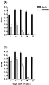
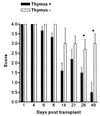
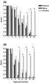
Oropharyngeal candidiasis is associated with defects in cell-mediated immunity and is commonly seen in human immunodeficiency virus positive individuals and AIDS patients. A model for oral candidiasis in T-cell-deficient BALB/c and CBA/CaH nu/nu mice was established. After inoculation with 10(8) Candida albicans yeasts, these mice displayed increased levels of oral colonization compared to euthymic control mice and developed a chronic oropharyngeal infection. Histopathological examination of nu/nu oral tissues revealed extensive hyphae penetrating the epithelium, with polymorphonuclear leukocyte microabscess formation. Adoptive transfer of either naive or immune lymphocytes into immunodeficient mice resulted in the recovery of these animals from the oral infection. Reconstitution of immunodeficient mice with naive CD4(+) but not CD8(+) T cells significantly decreased oral colonization compared to controls. Interleukin-12 and gamma interferon were detected in the draining lymph nodes of immunodeficient mice following reconstitution with naive lymphocytes. This study demonstrates the direct requirement for T lymphocytes in recovery from oral candidiasis and suggests that this is associated with the production of cytokines by CD4(+) T helper cells.
Figures


Similar articles
-
T cells augment monocyte and neutrophil function in host resistance against oropharyngeal candidiasis.Infect Immun. 2001 Oct;69(10):6110-8. doi: 10.1128/IAI.69.10.6110-6118.2001. Infect Immun. 2001. PMID: 11553549 Free PMC article.
-
Both CD4+ and CD8+ lymphocytes reduce the severity of tissue lesions in murine systemic cadidiasis, and CD4+ cells also demonstrate strain-specific immunopathological effects.Microbiology (Reading). 1999 Jul;145 ( Pt 7):1631-1640. doi: 10.1099/13500872-145-7-1631. Microbiology (Reading). 1999. PMID: 10439402
-
Cytokines in the oral mucosa of mice infected with Candida albicans.Oral Microbiol Immunol. 2002 Dec;17(6):375-8. doi: 10.1034/j.1399-302x.2002.170607.x. Oral Microbiol Immunol. 2002. PMID: 12485329
-
Candida-host interactions in HIV disease: relationships in oropharyngeal candidiasis.Adv Dent Res. 2006 Apr 1;19(1):80-4. doi: 10.1177/154407370601900116. Adv Dent Res. 2006. PMID: 16672555 Review.
-
Candida-host interactions in HIV disease: implications for oropharyngeal candidiasis.Adv Dent Res. 2011 Apr;23(1):45-9. doi: 10.1177/0022034511399284. Adv Dent Res. 2011. PMID: 21441480 Free PMC article. Review.
Cited by
-
Detection of a new embryonic antigen (ESA-10) in the blood of patients with cancer: preliminary results in the United States.Med Oncol. 2011 Mar;28(1):67-70. doi: 10.1007/s12032-010-9428-0. Epub 2010 Jan 27. Med Oncol. 2011. PMID: 20107933
-
Divergent EGFR/MAPK-Mediated Immune Responses to Clinical Candida Pathogens in Vulvovaginal Candidiasis.Front Immunol. 2022 May 26;13:894069. doi: 10.3389/fimmu.2022.894069. eCollection 2022. Front Immunol. 2022. PMID: 35720274 Free PMC article.
-
Recent mouse and rat methods for the study of experimental oral candidiasis.Virulence. 2013 Jul 1;4(5):391-9. doi: 10.4161/viru.25199. Epub 2013 May 28. Virulence. 2013. PMID: 23715031 Free PMC article. Review.
-
Immune cell-mediated protection against vaginal candidiasis: evidence for a major role of vaginal CD4(+) T cells and possible participation of other local lymphocyte effectors.Infect Immun. 2002 Sep;70(9):4791-7. doi: 10.1128/IAI.70.9.4791-4797.2002. Infect Immun. 2002. PMID: 12183521 Free PMC article.
-
Yeasts in the gut: from commensals to infectious agents.Dtsch Arztebl Int. 2009 Dec;106(51-52):837-42. doi: 10.3238/arztebl.2009.0837. Epub 2009 Dec 18. Dtsch Arztebl Int. 2009. PMID: 20062581 Free PMC article. Review.
References
-
- Ashman, R. B. 1998. A gene (Carg1) that regulates tissue resistance to Candida albicans maps to chromosome 14 of the mouse. Microb. Pathog. 25:333-335. - PubMed
-
- Ashman, R. B. 1997. Genetic determination of susceptibility and resistance in the pathogenesis of Candida albicans infection. FEMS Immunol. Med. Microbiol. 19:183-189. - PubMed
-
- Ashman, R. B. 1987. Mouse candidiasis. II. Host responses are T-cell dependent and regulated by genes in the major histocompatibility complex. Immunogenetics 25:200-203. - PubMed
-
- Ashman, R. B., E. M. Bolitho, and J. M. Papadimitriou. 1993. Patterns of resistance to Candida albicans in inbred mouse strains. Immunol. Cell. Biol. 71:221-225. - PubMed
-
- Ashman, R. B., A. Fulurija, and J. M. Papadimitriou. 1997. Evidence that two independent host genes influence the severity of tissue damage and susceptibility to acute pyelonephritis in murine systemic candidiasis. Microb. Pathog. 22:187-192. - PubMed
Publication types
MeSH terms
Substances
LinkOut - more resources
Full Text Sources
Medical
Research Materials

