Comparative Study
. 2002 Feb;70(2):878-88.
doi: 10.1128/IAI.70.2.878-888.2002.

Comparison of biofilms formed by Candida albicans and Candida parapsilosis on bioprosthetic surfaces

Affiliations
Comparative Study

Comparison of biofilms formed by Candida albicans and Candida parapsilosis on bioprosthetic surfaces

D M Kuhn et al. Infect Immun. 2002 Feb.

Abstract

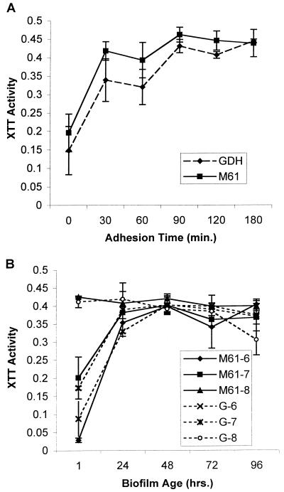
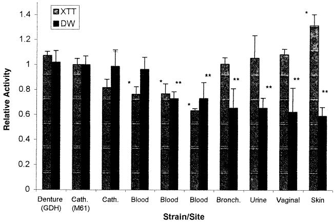
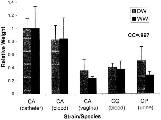
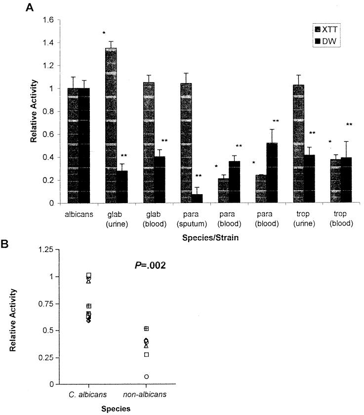
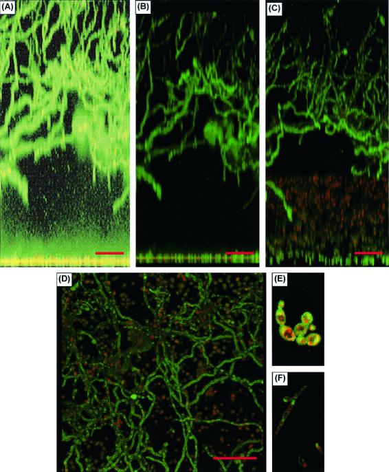
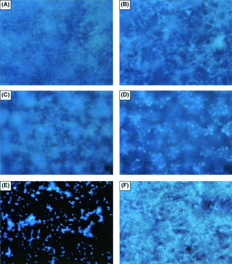
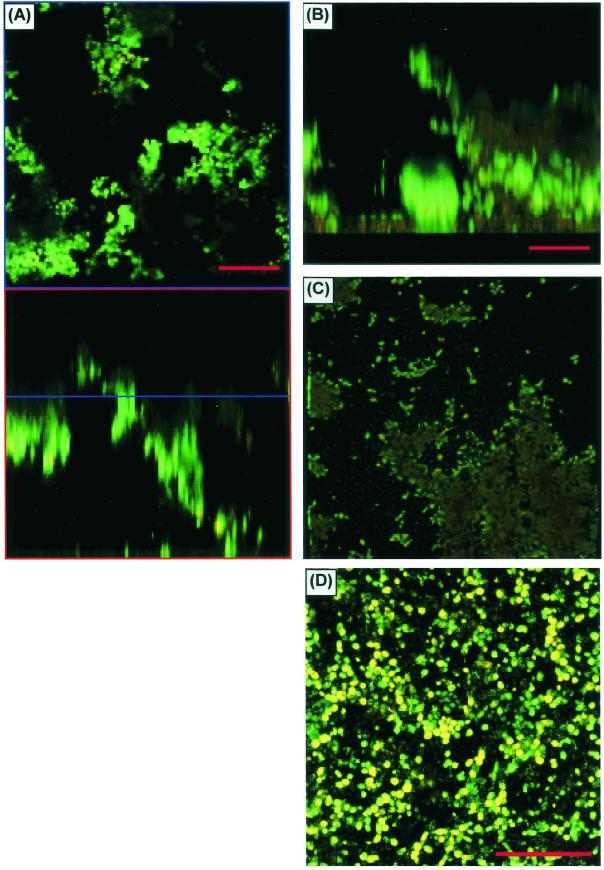
Little is known about fungal biofilms, which may cause infection and antibiotic resistance. In this study, biofilm formation by different Candida species, particularly Candida albicans and C. parapsilosis, was evaluated by using a clinically relevant model of Candida biofilm on medical devices. Candida biofilms were allowed to form on silicone elastomer and were quantified by tetrazolium (XTT) and dry weight (DW) assays. Formed biofilm was visualized by using fluorescence microscopy and confocal scanning laser microscopy with Calcofluor White (Sigma Chemical Co., St. Louis, Mo.), concanavalin A-Alexafluor 488 (Molecular Probes, Eugene, Oreg.), and FUN-1 (Molecular Probes) dyes. Although minimal variations in biofilm production among invasive C. albicans isolates were seen, significant differences between invasive and noninvasive isolates (P < 0.001) were noted. C. albicans isolates produced more biofilm than C. parapsilosis, C. glabrata, and C. tropicalis isolates, as determined by DW assays (P was <0.001 for all comparisons) and microscopy. Interestingly, noninvasive isolates demonstrated a higher level of XTT activity than invasive isolates. On microscopy, C. albicans biofilms had a morphology different from that of other species, consisting of a basal blastospore layer with a dense overlying matrix composed of exopolysaccharides and hyphae. In contrast, C. parapsilosis biofilms had less volume than C. albicans biofilms and were comprised exclusively of clumped blastospores. Unlike planktonically grown cells, Candida biofilms rapidly (within 6 h) developed fluconazole resistance (MIC, >128 microg/ml). Importantly, XTT and FUN-1 activity showed biofilm cells to be metabolically active. In conclusion, our data show that C. albicans produces quantitatively larger and qualitatively more complex biofilms than other species, in particular, C. parapsilosis.


Figures

FIG. 1.
FIG. 1.
Optimization of Candida biofilm growth on SE disks. (A) Effect of adhesion time on biofilm formation by C. albicans (strains M61 and GDH2346 [GDH]). XTT activity was measured at 492 nm. (B) Effect of C. albicans inoculum concentration on subsequent biofilm formation. The scheme for strain names is as follows: M61-7, C. albicans M61 at 107; G-8, GDH2346 at 108; and so on. Assays were performed in quadruplicate. Each result is representative of at least two experiments. All values are means and standard deviations.
FIG. 2.
FIG. 2.
Comparison of biofilms formed by different C. albicans isolates. The graph shows XTT and DW values for isolates of C. albicans obtained from different clinical sites. Cath., catheter; Bronch., bronchial site. Results were normalized to those for C. albicans strain M61. Assays were performed in quadruplicate. Each result is representative of at least two experiments. All values are means and standard deviations. Comparisons are significant for P values of <0.0011. A single asterisk indicates a P value of <0.001 for an XTT value of an isolate compared to M61; a double asterisk indicates a P value of <0.001 for a DW value of an isolate compared to M61.
FIG. 3.
FIG. 3.
Correlation of DW and WW measurements. The graph shows DW and WW measurements for different Candida isolates. CA, CG, and CP, C. albicans, C. glabrata, and C. parapsilosis, respectively. Results were normalized to those for C. albicans strain M61. Assays were performed in quadruplicate. Each result is representative of at least two experiments. All values are means and standard deviations. CC, correlation coefficient for the comparison of DW and WW measurements for all five isolates.
FIG. 4.
FIG. 4.
Comparison of biofilms formed by different Candida species. (A) XTT and DW measurements for different species of Candida. albicans, glab, para, and trop, C. albicans, C. glabrata, C. parapsilosis, and C. tropicalis, respectively. Results were normalized to those for C. albicans strain M61. Assays were performed in quadruplicate. Each result is representative of at least two experiments. All values are means and standard deviations. Comparisons are significant for P values of <0.0011. A single asterisk indicates a P value of <0.001 for an XTT value of an isolate compared to M61; a double asterisk indicates a P value of <0.001 for a DW value of an isolate compared to M61. (B) DW values for C. albicans versus non-C. albicans species shown in Fig. 2 and 4A, respectively.
FIG. 5.
FIG. 5.
CSLM examination of C. albicans biofilms. (A) CSLM image obtained with CAAF and FUN-1 staining and processed by a transparent projection technique to show a lateral view of the biofilm, which is ≈450 μm deep. A ×20 water immersion objective was used. Images were enhanced to highlight CAAF. The green CAAF staining highlights cell walls of the basal blastospore layer (bottom) and the upper hyphal layer. The diffuse staining of the extracellular space reflects CAAF binding matrix polysaccharides throughout the biofilm (the mild basilar enhancement is due to an out-of-focus artifact enhanced by the processing protocol). (B) Same lateral view but processed to show both CAAF and FUN-1 staining. (C) Tilt view (≈30°) illustrating red emission by converted FUN-1 in blastospores. (D) Top-down reconstructed view of the same image stack. (E and F) Representative red-enhanced image of the basal blastospore layer (E) and a similar image of hyphae in the upper matrix (F). Both panels E and F show FUN-1 conversion, as indicated by red dots within the cells.
FIG. 6.
FIG. 6.
FM examination of fungal biofilms formed by different Candida species. (A and B) FM examination of the basal and upper layers of a C. albicans biofilm, respectively. The biofilm was stained with Calcofluor White and viewed at ×10. (C and D) Similar views of C. parapsilosis, although the view of the upper layer is at a lower altitude than that in panel B, due to the limited thickness of the matrix. (E and F) Views of C. glabrata and C. tropicalis, respectively. Other samples of C. tropicalis and C. parapsilosis had the same appearance as that in panel E.
FIG. 7.
FIG. 7.
CSLM characterization of C. parapsilosis biofilms. (A) Horizontal (top) and vertical (bottom) slice reconstructions of a C. parapsilosis biofilm examined by CSLM with CAAF and FUN-1. Images were obtained with a ×20 water immersion objective. Green haze is an out-of-focus artifact, not extracellular matrix. (B) Image reconstructed by using transparent projection to show the lateral aspect, which is ≈117 μm deep. (C) Top-down view of the same image stack. (D) View of a different C. parapsilosis isolate, which failed to form appreciable matrix.

Similar articles

Cited by

References

    1. Anaissie, E. J., J. H. Rex, O. Uzon, and S. Vartivarian. 1998. Predictors of adverse outcome in cancer patients with candidemia. Am. J. Med. 104:238-245. - PubMed
    1. Baillie, G. S., and L. J. Douglas. 1999. Candida biofilms and their susceptibility to antifungal agents. Methods Enzymol. 310:644-656. - PubMed
    1. Baillie, G. S., and L. J. Douglas. 1999. Role of dimorphism in the development of Candida albicans biofilms. J. Med. Microbiol. 48:671-679. - PubMed
    1. Baillie, G. S., and L. J. Douglas. 2000. Matrix polymers of Candida biofilms and their possible role in biofilm resistance to antifungal agents. J. Antimicrob. Chemother. 46:397-403. - PubMed
    1. Banejee, S. N., G. T. Emori, D. H. Culver, R. P. Gaynes, W. R. Jarvis, T. Horan, J. R. Edwards, et al. 1991. Secular trends in nosocomial primary bloodstream infections in the United States, 1980-1989. Am. J. Med. 91(Suppl. 3B):86-89. - PubMed

Publication types

MeSH terms