. 2002 Feb;70(2):889-98.
doi: 10.1128/IAI.70.2.889-898.2002.

Rapid activation of protein tyrosine kinase and phospholipase C-gamma2 and increase in cytosolic free calcium are required by Ehrlichia chaffeensis for internalization and growth in THP-1 cells

Affiliations

Rapid activation of protein tyrosine kinase and phospholipase C-gamma2 and increase in cytosolic free calcium are required by Ehrlichia chaffeensis for internalization and growth in THP-1 cells

Mingqun Lin et al. Infect Immun. 2002 Feb.

Abstract

Ehrlichia chaffeensis, a bacterium that cannot survive outside the eukaryotic cell, proliferates exclusively in human monocytes and macrophages. In this study, signaling events required for ehrlichial infection of human monocytic cell line THP-1 were characterized. Entry and proliferation of E. chaffeensis in THP-1 cells were significantly blocked by various inhibitors that can regulate calcium signaling, including 8-(diethylamino)octyl-3,4,5-trimethoxybenzoate and 2-aminoethoxydiphenyl borate (intracellular calcium mobilization inhibitors), verapamil and 1-[beta-[3-(4-methoxyphenyl)propyl]-4-methoxyphenethyl]-1H-imidazole (SKF-96365) (calcium channel inhibitors), neomycin and 1-(6-[[17beta-3-methoxyestra-1,3,5(10)-trien-17-yl]amino]hexyl)-1H-pyrrole-2,5-dione (U-73122) (phospholipase C [PLC] inhibitors), monodansylcadaverine (a transglutaminase [TGase] inhibitor), and genistein (a protein tyrosine kinase [PTK] inhibitor). Addition of E. chaffeensis resulted in rapid increases in the level of inositol 1,4,5-trisphosphate (IP(3)) and the level of cytosolic free calcium ([Ca(2+)](i)) in THP-1 cells, which were prevented by pretreatment of THP-1 cells with inhibitors of TGase, PTK, and PLC. E. chaffeensis induced rapid tyrosine phosphorylation of PLC-gamma2, and the presence of a PLC-gamma2 antisense oligonucleotide in THP-1 cells significantly blocked ehrlichial infection. Furthermore, tyrosine-phosphorylated proteins and PLC-gamma2 were colocalized with ehrlichial inclusions, as determined by double-immunofluorescence labeling. The heat-sensitive component of viable E. chaffeensis cells was essential for these signaling events. E. chaffeensis, therefore, can recruit interacting signal-transducing molecules and induce the following signaling events required for the establishment of infection in host cells: protein cross-linking by TGase, tyrosine phosphorylation, PLC-gamma2 activation, IP(3) production, and an increase in [Ca(2+)](i).


Figures
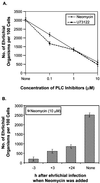
FIG. 1.
FIG. 1.
Effects of PLC inhibitors on E. chaffeensis infection. The PLC inhibitors neomycin and U-73122 blocked E. chaffeensis infection in a dose-dependent manner (neomycin and U-73122 were added 1 h prior to ehrlichial infection) (A) or in a time-dependent manner (10 μM neomycin was added to THP-1 cells before or after infection) (B). Infectivity was determined 3 days p.i. as described in Materials and Methods. The values are means ± standard deviations (n = 3) and are representative of the results of more than three independent experiments.
FIG. 2.
FIG. 2.
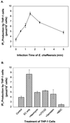
Effects of ehrlichial infection and inhibitors on production of IP3 in THP-1 cells. (A) Rapid release of IP3 induced by ehrlichial infection. The release reached a peak (approximately threefold) at 1.5 min p.i. and decreased to the background level within 5 min. (B) Release of IP3 induced by E. chaffeensis (EC) prevented by pretreatment with inhibitors of PTK, PLC, and TGase. Cells were pretreated with or without inhibitors 1 h before ehrlichial infection, and IP3 samples were extracted with perchloric acid at different times (A) or 1.5 min p.i. (peak release of IP3) (B). The release of IP3 was determined by measuring competitive inhibition of binding of 3H-labeled IP3 to IP3-binding protein. The values are means ± standard deviations (n = 3) and are representative of the results of three independent experiments.
FIG. 3.
FIG. 3.
Effects of ehrlichial infection and inhibitors on [Ca2+]i in a population of cells. [Ca2+]i increased rapidly and transiently in THP-1 cells infected with E. chaffeensis (E.C.) (A), but the increase was prevented by pretreatment with inhibitors of calcium mobilization (verapamil and TMB-8) (B) or inhibitors of PLC (neomycin), PTK (genistein), and TGase (MDC) (C). The intracellular calcium levels in fluo-3-loaded THP-1 cells were determined with a fluorometer as described in Materials and Methods. The inhibitors were added to cells 1 h before the assay, and host-cell-free E. chaffeensis was added to THP-1 cells at the time indicated by the vertical arrow (approximately 50 s p.i.). Assays were carried out at 30°C in a circulating water bath. Fluorescence was measured at 525 nm at 5-s intervals, and Kd was calibrated by using a calcium calibration kit from Molecular Probes. The values are representative of the results of at least four independent experiments. moi, multiplicity of infection.
FIG. 4.
FIG. 4.
Effects of ehrlichial infection and inhibitors on [Ca2+]i in an individual cell. The rapid and transient increase in [Ca2+]i (as shown by the ratio of emission by fura-2 at 520 nm under stimulation at 340 nm to emission by fura-2 at 520 nm under stimulation at 380 nm) was induced by E. chaffeensis (E.C.) infection in an individual THP-1 cell (A). The increase in [Ca2+]i required active participation of live E. chaffeensis since heat-treated (42 or 60°C, 15 min) E. chaffeensis did not increase the [Ca2+]i. However, the increase in [Ca2+]i induced by ehrlichial infection was prevented by the calcium mobilization inhibitors SKF-96365 and 2-APB (B) or by the PLC inhibitor neomycin and the PTK inhibitor genistein (C). THP-1 cells were loaded with fura-2/AM and treated with or without inhibitors for 1 h before measurements were obtained. Cells were added to poly-l-lysine-coated coverslip chambers, and images (emission at 520 nm after excitation at 340 and 380 nm) were recorded at 1-s intervals for at least 5 min. Host-cell-free E. chaffeensis was added to the chambers after measurements had been obtained for 30 s (vertical arrow). The values are representative of the results of at least three independent experiments, and similar results were obtained for more than 20 cells in each experiment.
FIG. 5.
FIG. 5.
Tyrosine phosphorylation of PLC-γ2 in ehrlichial infection. (A) A 145-kDa protein was rapidly tyrosine phosphorylated in THP-1 cells upon binding of E. chaffeensis (EC). This protein was confirmed to be PLC-γ2 by immunoprecipitation (IP) with anti-PLC-γ2 antibody and by Western blot (WB) analysis with detection with anti-phosphotyrosine (pTyr) antibody (B) or anti-PLC-γ2 antibody (C). Tyrosine phosphorylation of PLC-γ2 was reduced when E. chaffeensis was pretreated at 42 or 60°C for 15 min (D). THP-1 cells were pretreated with genistein (Gen) or MDC for 1 h prior to the addition of E. chaffeensis. At different times, cells were lysed in RIPA buffer. Whole-cell lysates were immunoprecipitated with anti-PLC-γ2 antibody, and the immunoprecipitates or whole-cell lysates were subjected to Western blotting by using anti-phosphotyrosine antibody or anti-PLC-γ2 antibody. The results are representative of the results of more than four independent experiments in which similar results were obtained. CTL, control.
FIG. 6.
FIG. 6.
Colocalization of E. chaffeensis inclusions with PLC-γ2 and tyrosine-phosphorylated proteins but not with PLC-γ1. E. chaffeensis-infected THP-1 cells (2 days p.i.) were double labeled as described in the text and observed by confocal microscopy. The following antibodies were labeled: E. chaffeensis (green, left panels) and PLC-γ1 or -γ2 or phosphotyrosine (red, middle panels). The panels on the right are superimposed images viewed with green and red filters. Note the colocalization of E. chaffeensis with PLC-γ2 and phosphotyrosine (yellow). The results are representative of the results of three independent labeling experiments.
FIG. 7.
FIG. 7.
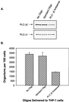
Effect of PLC-γ2 antisense oligonucleotides on E. chaffeensis infection. (A) Delivery of PLC-γ2 antisense oligonucleotides (Oligo) into THP-1 cells significantly reduced the level of PLC-γ2 but not the level of PLC-γ1 compared to the levels in untransfected cells or cells transfected with standard control oligonucleotides, as determined by Western blotting. (B) Delivery of PLC-γ2 antisense oligonucleotides into THP-1 cells also significantly reduced E. chaffeensis infection (>50%) (P < 0.05). THP-1 cells were transfected with anti-PLC-γ2 oligonucleotides or standard control oligonucleotides as described in Materials and Methods. Three days after the delivery of antisense oligonucleotides, the cells were infected with host-cell-free E. chaffeensis. On 3 day p.i., infectivity was determined, cells were extracted by using RIPA buffer, and a Western blot analysis was carried out to determine the levels of PLC-γ1 and PLC-γ2. The results are representative of the results of three independent experiments.
FIG. 8.
FIG. 8.
Model of signaling pathway induced by E. chaffeensis infection. Binding of E. chaffeensis to its receptor(s) on host monocytes triggers protein cross-linking by TGase, which activates the receptor or nonreceptor tyrosine kinase. PTK activation mediates the translocation and tyrosine phosphorylation of PLC-γ2, which produces IP3. IP3 in turn induces Ca2+ release from internal stores. The depletion of Ca2+ from internal stores activates store-operated calcium channels on the plasma membrane, and Ca2+ influx from external spaces contributes to the bulk of the increase in [Ca2+]i in response to the infection. Activation of TGase by Ca2+ constitutes a positive feedback loop, and this may facilitate entry of ehrlichiae into host cells or ehrlichial infection. The dotted lines indicate suggested pathways, and the solid lines indicate proven pathways.

Similar articles

Cited by

References

    1. Abe, S., K. Yamashita, H. Kohno, and Y. Ohkubo. 2000. Involvement of transglutaminase in the receptor-mediated endocytosis of mouse peritoneal macrophages. Biol. Pharm. Bull. 23:1511–1513. - PubMed
    1. Anderson, C. L., L. Shen, D. M. Eicher, M. D. Wewers, and J. K. Gill. 1990. Phagocytosis mediated by three distinct Fc gamma receptor classes on human leukocytes. J. Exp. Med. 171:1333–1345. - PMC - PubMed
    1. Andersson, S. G., A. Zomorodipour, J. O. Andersson, T. Sicheritz-Ponten, U. C. Alsmark, R. M. Podowski, A. K. Naslund, A. S. Eriksson, H. H. Winkler, and C. G. Kurland. 1998. The genome sequence of Rickettsia prowazekii and the origin of mitochondria. Nature 396:133–140. - PubMed
    1. Bain, C., R. Keller, G. K. Collington, L. R. Trabulsi, and S. Knutton. 1998. Increased levels of intracellular calcium are not required for the formation of attaching and effacing lesions by enteropathogenic and enterohemorrhagic Escherichia coli. Infect. Immun. 66:3900–3908. - PMC - PubMed
    1. Baldwin, T. J., W. Ward, A. Aitken, S. Knutton, and P. H. Williams. 1991. Elevation of intracellular free calcium levels in HEp-2 cells infected with enteropathogenic Escherichia coli. Infect. Immun. 59:1599–1604. - PMC - PubMed

Publication types

LinkOut - more resources