

Cell differentiation is a key determinant of cathelicidin LL-37/human cationic antimicrobial protein 18 expression by human colon epithelium
- PMID: 11796631
- PMCID: PMC127717
- DOI: 10.1128/IAI.70.2.953-963.2002
Cell differentiation is a key determinant of cathelicidin LL-37/human cationic antimicrobial protein 18 expression by human colon epithelium
Abstract
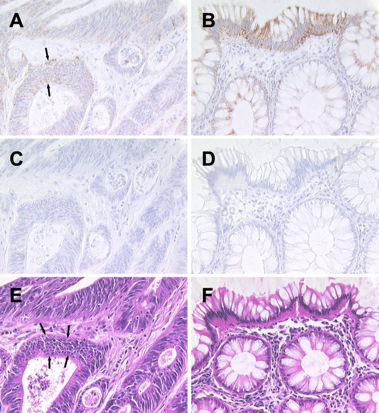
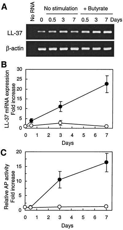
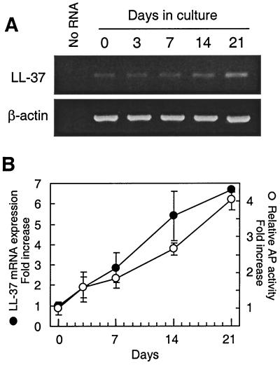
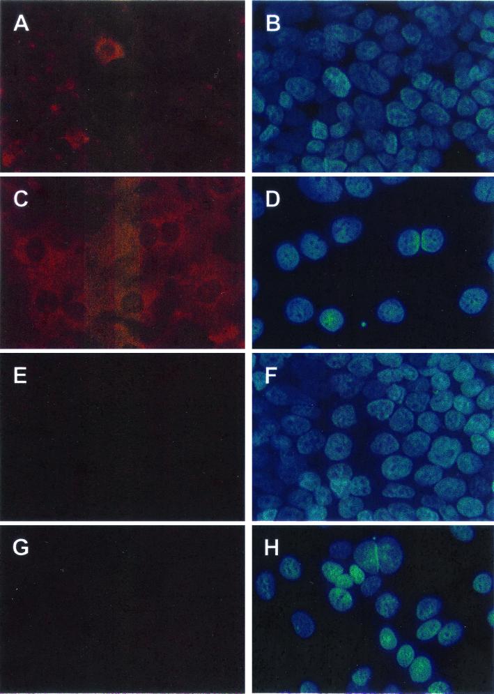
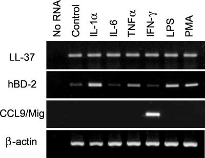
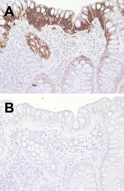
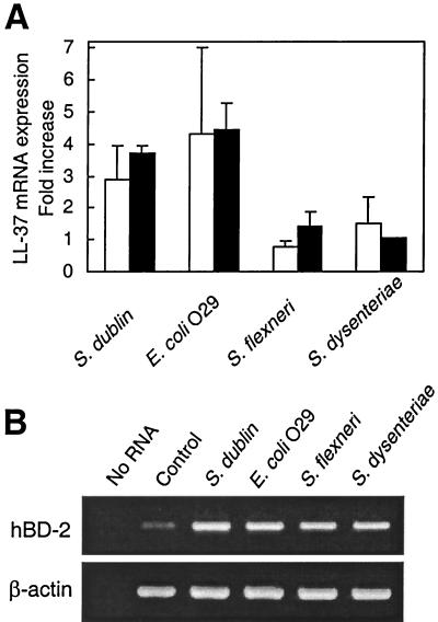
Antimicrobial peptides are highly conserved evolutionarily and are thought to play an important role in innate immunity at intestinal mucosal surfaces. To better understand the role of the antimicrobial peptide human cathelicidin LL-37/human cationic antimicrobial protein 18 (hCAP18) in intestinal mucosal defense, we characterized the regulated expression and production of this peptide by human intestinal epithelium. LL-37/hCAP18 is shown to be expressed within epithelial cells located at the surface and upper crypts of normal human colon. Little or no expression was seen within the deeper colon crypts or within epithelial cells of the small intestine. Paralleling its expression in more differentiated epithelial cells in vivo, LL-37/hCAP18 mRNA and protein expression was upregulated in spontaneously differentiating Caco-2 human colon epithelial cells and in HCA-7 human colon epithelial cells treated with the cell differentiation-inducing agent sodium butyrate. LL-37/hCAP18 expression by colon epithelium does not require commensal bacteria, since LL-37/hCAP18 is produced with a similar expression pattern by epithelial cells in human colon xenografts that lack a luminal microflora. LL-37/hCAP18 mRNA was not upregulated in response to tumor necrosis factor alpha, interleukin 1alpha (IL-1alpha), gamma interferon, lipopolysaccharide, or IL-6, nor did the expression patterns and levels of LL-37/hCAP18 in the epithelium of the normal and inflamed colon differ. On the other hand, infection of HCA-7 cells with Salmonella enterica serovar Dublin or enteroinvasive Escherichia coli modestly upregulated LL-37/hCAP18 mRNA expression. We conclude that differentiated human colon epithelium expresses LL-37/hCAP18 as part of its repertoire of innate defense molecules and that the distribution and regulated expression of LL-37/hCAP18 in the colon differs markedly from that of other enteric antimicrobial peptides, such as defensins.
Figures

Similar articles
-
Heterogeneous expression of human cathelicidin hCAP18/LL-37 in inflammatory bowel diseases.Eur J Gastroenterol Hepatol. 2006 Jun;18(6):615-21. doi: 10.1097/00042737-200606000-00007. Eur J Gastroenterol Hepatol. 2006. PMID: 16702850
-
Expression of LL-37 by human gastric epithelial cells as a potential host defense mechanism against Helicobacter pylori.Gastroenterology. 2003 Dec;125(6):1613-25. doi: 10.1053/j.gastro.2003.08.028. Gastroenterology. 2003. PMID: 14724813
-
Expression and regulation of the human beta-defensins hBD-1 and hBD-2 in intestinal epithelium.J Immunol. 1999 Dec 15;163(12):6718-24. J Immunol. 1999. PMID: 10586069
-
The human cathelicidin hCAP18/LL-37: a multifunctional peptide involved in mycobacterial infections.Peptides. 2010 Sep;31(9):1791-8. doi: 10.1016/j.peptides.2010.06.016. Epub 2010 Jun 25. Peptides. 2010. PMID: 20600427 Review.
-
Development, validation and implementation of an in vitro model for the study of metabolic and immune function in normal and inflamed human colonic epithelium.Dan Med J. 2015 Jan;62(1):B4973. Dan Med J. 2015. PMID: 25557335 Review.
Cited by
-
Ethanol-induced changes to the gut microbiome compromise the intestinal homeostasis: a review.Gut Microbes. 2024 Jan-Dec;16(1):2393272. doi: 10.1080/19490976.2024.2393272. Epub 2024 Sep 3. Gut Microbes. 2024. PMID: 39224006 Free PMC article. Review.
-
MicroRNA-orchestrated pathophysiologic control in gut homeostasis and inflammation.BMB Rep. 2016 May;49(5):263-9. doi: 10.5483/bmbrep.2016.49.5.041. BMB Rep. 2016. PMID: 26923304 Free PMC article. Review.
-
Intestinal innate immunity and the pathogenesis of Salmonella enteritis.Immunol Res. 2007;37(1):61-78. doi: 10.1007/BF02686090. Immunol Res. 2007. PMID: 17496347 Free PMC article.
-
Intestinal antimicrobial peptides during homeostasis, infection, and disease.Front Immunol. 2012 Oct 9;3:310. doi: 10.3389/fimmu.2012.00310. eCollection 2012. Front Immunol. 2012. PMID: 23087688 Free PMC article.
-
Cathelicidin-mediated lipopolysaccharide signaling via intracellular TLR4 in colonic epithelial cells evokes CXCL8 production.Gut Microbes. 2020 Nov 9;12(1):1785802. doi: 10.1080/19490976.2020.1785802. Epub 2020 Jul 13. Gut Microbes. 2020. PMID: 32658599 Free PMC article.
References
-
- Agerberth, B., J. Charo, J. Werr, B. Olsson, F. Idali, L. Lindbom, R. Kiessling, H. Jörnvall, H. Wigzell, and G. H. Gudmundsson. 2000. The human antimicrobial and chemotactic peptides LL-37 and α-defensins are expressed by specific lymphocyte and monocyte populations. Blood 96:3086-3093. - PubMed
-
- Ayabe, T., D. P. Satchell, C. L. Wilson, W. C. Parks, M. E. Selsted, and A. J. Ouellette. 2000. Secretion of microbicidal α-defensins by intestinal Paneth cells in response to bacteria. Nat. Immunol. 1:113-118. - PubMed
-
- Bernet-Camard, M. F., M. H. Coconnier, S. Hudault, and A. L. Servin. 1996. Differentiation-associated antimicrobial functions in human colon adenocarcinoma cell lines. Exp. Cell Res. 226:80-89. - PubMed
Publication types
MeSH terms
Substances
Grants and funding
LinkOut - more resources
Full Text Sources
Other Literature Sources
