. 2002 Jan 15;16(2):183-97.
doi: 10.1101/gad.959402.

The mitotic spindle is required for loading of the DASH complex onto the kinetochore

Affiliations

The mitotic spindle is required for loading of the DASH complex onto the kinetochore

Yumei Li et al. Genes Dev. .

Abstract

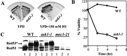
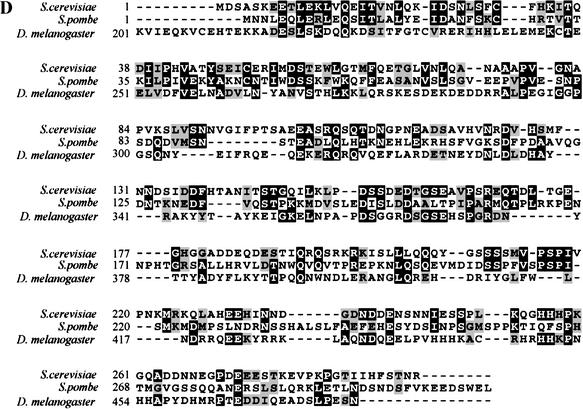
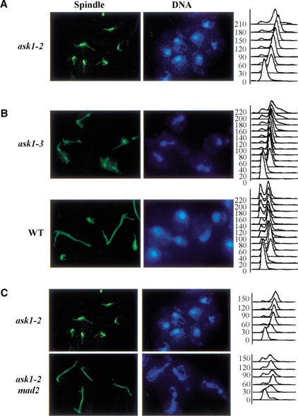
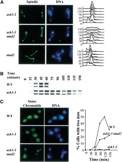
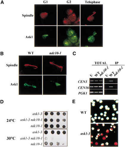
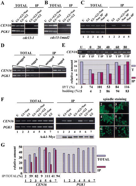
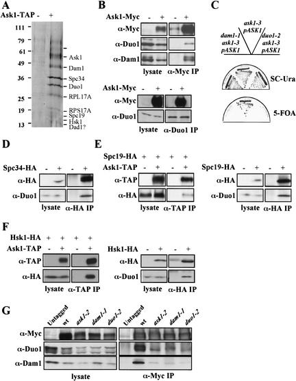
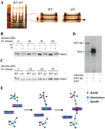
A role for the mitotic spindle in the maturation of the kinetochore has not been defined previously. Here we describe the isolation of a novel and conserved essential gene, ASK1, from Saccharomyces cerevisiae involved in this process. ask1 mutants display either G(2)/M arrest or segregation of DNA masses without the separation of sister chromatids, resulting in massive nondisjunction and broken spindles. Ask1 localizes along mitotic spindles and to kinetochores, and cross-links to centromeric DNA. Microtubules are required for Ask1 binding to kinetochores, and are partially required to maintain its association. We found Ask1 is part of a multisubunit complex, DASH, that contains approximately 10 components, including several proteins essential for mitosis including Dam1, Duo1, Spc34, Spc19, and Hsk1. The Ipl1 kinase controls the phosphorylation of Dam1 in the DASH complex and may regulate its function. We propose that DASH is a microtubule-binding complex that is transferred to the kinetochore prior to mitosis, thereby defining a new step in kinetochore maturation.


Figures

Figure 1
Figure 1
Characterization of ask1-1 mutant cells. (A) ask1-1 mutants are HU sensitive. Wild-type (Y300) and ask1-1 (Y928) mutants were streaked on YPD plates containing 150 mM HU at 30°C and incubated for 3 d. (B) ask1-1 loses viability in HU. α-factor arrested wild-type (Y300) and ask1-1 (Y928) mutant cells were released into YPD containing 200 mM HU for the indicated times and plated on YPD at 30°C to determine survival. (C) Proper regulation of Rad53 in ask1-1 mutants. Log-phase cells of wild-type (Y300), ask1-1 (Y928), and mec1-21 (Y604) were treated with 200 mM HU (lane 2) or 0.5% MMS (lane 3) for 1.5 h at 30°C. Protein extracts were prepared, separated by SDS/PAGE, and immunoblotted with antibodies against Rad53. (Lane 1) Untreated control. (D) Alignment of S. cerevisiae Ask1 protein with its homologs from S. pombe and D. melanogaster. Identical residues are highlighted in black and conserved residues in gray.
Figure 1
Figure 1
Characterization of ask1-1 mutant cells. (A) ask1-1 mutants are HU sensitive. Wild-type (Y300) and ask1-1 (Y928) mutants were streaked on YPD plates containing 150 mM HU at 30°C and incubated for 3 d. (B) ask1-1 loses viability in HU. α-factor arrested wild-type (Y300) and ask1-1 (Y928) mutant cells were released into YPD containing 200 mM HU for the indicated times and plated on YPD at 30°C to determine survival. (C) Proper regulation of Rad53 in ask1-1 mutants. Log-phase cells of wild-type (Y300), ask1-1 (Y928), and mec1-21 (Y604) were treated with 200 mM HU (lane 2) or 0.5% MMS (lane 3) for 1.5 h at 30°C. Protein extracts were prepared, separated by SDS/PAGE, and immunoblotted with antibodies against Rad53. (Lane 1) Untreated control. (D) Alignment of S. cerevisiae Ask1 protein with its homologs from S. pombe and D. melanogaster. Identical residues are highlighted in black and conserved residues in gray.
Figure 2
Figure 2
Characterization of temperature-sensitive alleles of ASK1. (A) ask1-2 mutant cells arrest before anaphase entry. ask1-2 mutant cells (Y1104) were synchronized by α-factor and released into YPD at 37°C. (Left) The DNA and spindle morphology of ask1-2 cells at 90 min following the release. (Right) The DNA content determined by FACS analysis. (B) ask1-3 mutants display unusual spindle phenotypes at the restrictive temperature. Wild-type (Y300) and ask1-3 (Y1103) mutant cells were synchronized by α-factor and released at 35°C. At the indicated times after the release, aliquots were withdrawn to examine viability, DNA content, and nuclear spindle morphology. Photomicrographs show wild-type and ask1-3 mutant cells at 90 min after release. (C) ask1-2 (Y1104) and ask1-2 mad2 (Y1106) mutant cells were synchronized and released as in A. Aliquots were collected for FACS analysis, DNA, and spindle staining at the indicated times. Photomicrographs were taken of cells at 90 min after the release.
Figure 3
Figure 3
ask1-3 mutants undergo massive nondisjunction and segregate chromosomes without the separation of sister chromatids. (A) The spindle checkpoint is active in ask1-3 mutants. ask1-3 (Y1103), ask1-3 mad2 (Y1107), and mad2 (Y1101) mutants were arrested in G1 with α-factor and then released at 35°C. Ninety minutes after the release, samples were collected and stained with DAPI (DNA) and anti-tubulin antibodies (spindles). (B) Pds1 is stabilized in ask1-3 mutants. Wild-type (Y998) and ask1-3 mutant (Y1108) cells carrying the PDS1 gene fused to 18 copies of the myc epitope were synchronized with α-factor at 24°C and then released at 35°C. Cells were harvested every 15 min and processed for protein preparation. Pds1 levels were determined by Western blotting using anti-Myc antibodies. (C) Analysis of sister-chromatid separation in arrested ask1-3 mutant cells. Wild-type (Y974), ask1-3 mutant (Y1109), and ask1-3 mad2 (Y1110) cells containing a Tet–GFP fusion and tandem repeats of the Tet operator integrated at the URA3 locus 35 kb away from centromere on the left arm of ChrV were synchronized by α-factor at 24°C and then released at 35°C. Ninety minutes after release, samples were collected to examine sister chromatid separation by immunofluorescence microscopy.
Figure 4
Figure 4
Ask1 is a centromere binding protein. (A) Ask1 localizes to the mitotic spindle and spindle-pole bodies. Cells carrying an integrated ASK1–GFP gene (Y1111) were synchronized in G1 with α-factor and released at 30°C. Samples at different stages of the cell cycle were fixed with 5% formaldehyde for 30 min before immunofluorescence staining. (B) Localization of Ask1 in wild-type and ndc10-1 mutants. G1-arrested wild-type (Y1111) and ndc10-1 (Y1112) cells containing Ask1–GFP were released into 35°C for immunofluorescence microscopy. Representative telophase cells are shown. (C) Chromatin immunoprecipitation analysis (ChIP) of Ask1–Myc in wild-type (Y1113) and ndc10-1 (Y1115) cells. Untagged wild-type (Y300) cells (U) are included as control. Log-phase cultures were shifted to 35°C for 3 h before collecting samples. (D) Synthetic growth defects in ask1-3 ndc10-1 double mutants (Y1120). Tenfold dilutions of cells were spotted on plates incubated at 24°C and 30°C. (E) ask1-3 mutants exhibit elevated rates of chromosome loss. Wild-type (Y1117) or ask1-3 mutant (Y1118) strains harboring CFIII were grown in SC-uracil medium at 24°C and then plated out on YPD plates at 32°C and incubated for 4 d. Red sectors represent chromosome loss events.
Figure 5
Figure 5
The association of Ask1 to the centromere requires intact microtubules. (A) Ask1 centromeric binding is inhibited by nocodazole. α-factor-arrested cdc13-1 ASK1–MYC (Y1114) cells were released into prewarmed YPD with (G1–G2 + Noc) or without (G1–G2) nocodazole at 32°C. Seventy minutes later, samples were collected for ChIP analysis. Untagged wild-type cells are included as a control (U). (B) Depolymerization of microtubules, not the activation of the spindle checkpoint, blocks the loading of Ask1 to centromeric DNA. G1-arrested cdc13-1 mad2 (Y1116) cells containing Ask1–Myc were released into medium containing nocodazole for 70 min and processed for ChIP analysis as above. Untagged wild-type cells are included as a control (U). (C) The nocodazole-dependent centromere binding of Ask1 is reversible. α-factor-arrested cdc13-1 ASK1–MYC (Y1114) cells (G1) were released into prewarmed YPD with nocodazole at 32°C (G1–G2 + Noc). Seventy minutes later, cells were spun down and resuspended in fresh YPD at 32°C to remove nocodazole. Samples were collected at 30-min intervals for ChIP analysis (−Noc 30', −Noc 60'). Untagged wild-type cells are included as a control (U). (D) Control showing that the PCR reactions are in the linear range. Twofold serial dilutions of the precipitated and input samples used in lanes 1 and 7 in F were subjected to PCR. (E) The centromere binding of Ask1 in the cell cycle. ASK1–MYC (Y1113) cells were arrested at G1 with α-factor and then released at 30°C. Samples were collected every 20 min for ChIP analysis. DNAs from total (T) and immunoprecipitaed (IP) samples were analyzed and the PCR products were quantitated using NIH image software. Percent budding for the samples is shown below. (F) The centromere binding of Ask1 in G1 and G2-arrested cells is less sensitive to nocodazole treatment. cdc13-1 ASK1–MYC (Y1114) cells were arrested in G1 with α-factor or in G2 by incubating at 32°C before nocodazole was added. Seventy minutes later, samples were collected for ChIP analysis. Cells that were released from G1 to G2 were included as a control (lanes 4,5). (Bottom) The amounts of Ask1–Myc protein immunoprecipitated as determined by Western analysis. (Right) The control showing that nocodazole treatment destabilized microtubules. Shown here are anti-tubulin immunoflourescence images of cells used to prepare the DNA used in lanes 2–7. (G) Quantification of the PCR reactions in F. PCR products in F were run on a 3% agarose gel, visualized with ethidium bromide staining, and scanned for quantitation using NIH Image software.
Figure 6
Figure 6
The identification of DASH complex. (A) TAP-purified protein extracts from 2L of yeast culture from either ASK1–TAP (Y1173) or untagged strains (Y300) were resolved on 4%–20% SDS–polyacrylamide gels and stained with Coomassie blue. Protein bands were cut out and identified by mass spectrometry as described previously (Wang et al. 2000). Question marks indicate proteins whose identities are suspected but not yet determined. (B) Coimmunoprecipitation of Dam1 and Duo1 with Ask1–Myc. (Top) Ask1–Myc was immunoprecipitated with anti-Myc antibodies. Proteins from total protein extracts (lysate) or from the immunoprecipitated fractions (IP) were analyzed by immunoblotting, using antibodies against Dam1 and Duo1. (Bottom) The same extracts as above were immunoprecipitated with anti-Duo1 antibodies and Western blots were probed with anti-Myc antibodies. (C) ask1-3 is synthetically lethal with dam1-1 and duo1-2. Both ask1-3 dam1-1 (Y1171) and ask1-3 duo1-2 (Y1172) containing pJBN81 (ASK1–URA3) were struck upon SC-Ura plates or SC plates containing 5-FOA. (D) Coimmunoprecipitation of Duo1 with Spc34. Immunoprecipitation was carried out with anti-HA antibodies and Western blots were probed with anti-Duo1 antibodies. (E) Spc19 is a component of the DASH complex. (Left) Extracts from SPC19-HA and SPC19-HA ASK–-TAP strains were immunoprecipitated with anti-TAP antibodies (PAP: peroxidase-anti-peroxidase) and Western blots were probed with anti-TAP and anti-HA antibodies. (Right) Extracts from SPC19-HA and nontagged strains were immunoprecipitated with anti-HA antibodies and Western blots were probed with anti-HA and anti-Duo1 antibodies as indicated. (F) Hsk1 is a component of the DASH complex. (Left) Extracts from HSK1-HA and HSK1-HA ASK1-TAP strains were immunoprecipitated with anti-TAP antibodies (PAP) and Western blots were probed with anti-TAP and anti-HA antibodies. (Right) Extracts from HSK1-HA and nontagged strains were immunoprecipitated with anti-HA antibodies and Western blots were probed with anti-HA and anti-Duo1 antibodies. (G) ASK1, DAM1, and DUO1 are required for the formation of the DASH complex. Immunoprecipitations were carried out on extracts from ask1-2–MYC (Y1174), dam1-1 ASK1–MYC (Y1176), or duo1-2 ASK1–MYC (Y1175) strains and probed for Dam1, Duo1, and Ask1–Myc.
Figure 7
Figure 7
Ipl1 controls Dam1 phosphorylation. (A) The mobility of DASH complex proteins is altered in ipl1 mutants. TAP-purified protein extracts from 2 L of yeast culture from either ASK1–TAP (Y1173) or ASK1–TAP ipl1-321 mutants (Y1182) were resolved on 4%–20% SDS–polyacrylamide gel and silver stained. An expanded section is shown to illustrate the absence of certain bands in an ilp1 mutant strain. (B) Dam1 is a phosphoprotein. Dam1–Myc was prepared from extracts of Y1183 and treated with alkaline phosphatase in the presence or absence of phosphatase inhibitors. Proteins were then immunoblotted and probed with anti-Myc antibodies. (C) Ipl1 controls the phosphorylation of Dam1 in vivo. Protein extracts were prepared from Y1183 (DAM1–MYC) and Y1184 (DAM1–MYC ipl1-321) cells synchronized in G1 with α-factor and released into the cell cycle for the indicated time, and were then immunoblotted and probed with anti-MYC antibodies. (D) Ipl1 directly phosphorylates Dam1. Kinase assays were performed using GST–Ipl1 kinase (lanes 1,2) or GST alone as a control (lane 3). The substrate tested was the DASH complex purified from ipl1-321 ASK1–TAP (Y1182). The autophosphorylation of GST–Ipl1 and the phosphorylation of Dam1 are indicated at left. (E) Hypothetical model depicting the microtubule-dependent loading of the DASH complex to the kinetochore.

References

    1. Alcasabas AA, Osborn AJ, Bachant J, Hu F, Werler PJH, Bousset K, Kanji Furuya K, Diffley JFX, Carr A, Elledge SJ. Mrc1 transduces DNA replication stress signals to activate Rad53. Nat Cell Biol. 2001;3:958–965. - PubMed
    1. Amon A. The spindle checkpoint. Curr Opin Genet Dev. 1999;9:69–75. - PubMed
    1. Aparicio OM, Weinstein DM, Bell SP. Components and dynamics of DNA replication complexes in S. cerevisiae: Redistribution of MCM proteins and Cdc45p during S phase. Cell. 1997;91:59–69. - PubMed
    1. Biggins S, Murray AW. The budding yeast protein kinase Ipl1/Aurora allows the absence of tension to activate the spindle checkpoint. Genes & Dev. 2001;15:3118–3129. - PMC - PubMed
    1. Biggins S, Severin FF, Bhalla N, Sassoon I, Hyman AA, Murray AW. The conserved protein kinase Ipl1 regulates microtubule binding to kinetochores in budding yeast. Genes & Dev. 1999;13:532–544. - PMC - PubMed

Publication types

MeSH terms