. 2002 Jan 15;16(2):245-56.
doi: 10.1101/gad.949802.

E2F integrates cell cycle progression with DNA repair, replication, and G(2)/M checkpoints

Affiliations

E2F integrates cell cycle progression with DNA repair, replication, and G(2)/M checkpoints

Bing Ren et al. Genes Dev. .

Abstract

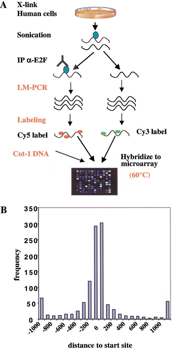
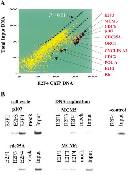
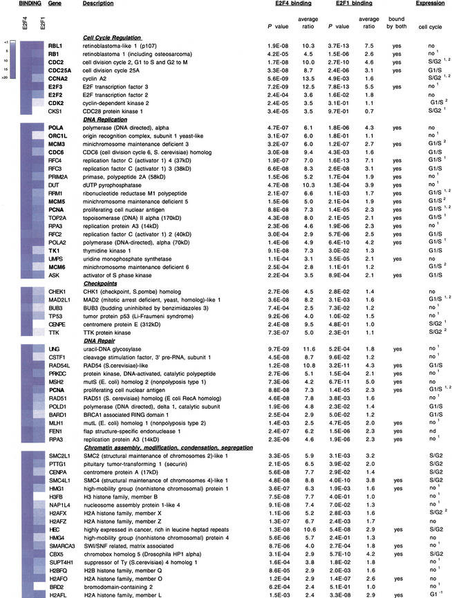
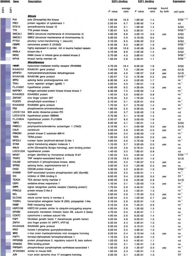
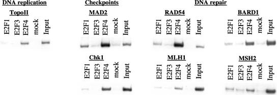
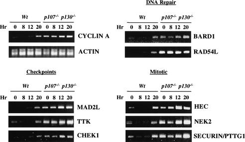
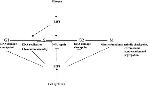
The E2F transcription factor family is known to play a key role in the timely expression of genes required for cell cycle progression and proliferation, but only a few E2F target genes have been identified. We explored the possibility that E2F regulators play a broader role by identifying additional genes bound by E2F in living human cells. A protocol was developed to identify genomic binding sites for DNA-binding factors in mammalian cells that combines immunoprecipitation of cross-linked protein-DNA complexes with DNA microarray analysis. Among approximately 1200 genes expressed during cell cycle entry, we found that the promoters of 127 were bound by the E2F4 transcription factor in primary fibroblasts. A subset of these targets was also bound by E2F1. Most previously identified target genes known to have roles in DNA replication and cell cycle control and represented on the microarray were confirmed by this analysis. We also identified a remarkable cadre of genes with no previous connection to E2F regulation, including genes that encode components of the DNA damage checkpoint and repair pathways, as well as factors involved in chromatin assembly/condensation, chromosome segregation, and the mitotic spindle checkpoint. Our data indicate that E2F directly links cell cycle progression with the coordinate regulation of genes essential for both the synthesis of DNA as well as its surveillance.


Figures

Figure 1
Figure 1
Strategy for mammalian factor location analysis using the 1.5K DNA microarray. (A) An illustration of the experimental protocol. Modifications to previous yeast protocols (Ren et al. 2000) are indicated in red. Primary human cells (WI-38) were cross-linked and sonicated, and chromatin was immunoprecipitated with anti-E2F antibodies. The resulting ChIP-enriched DNA was purified, amplified by LM-PCR together with total genomic DNA, labeled with the Cy5 and Cy3 fluorophores using random priming, and hybridized to the 1.5K microarray in the presence of Cot-1 DNA to reduce nonspecific hybridization. (B) The frequency and distribution of transcription factors binding to human promoters. The distance of transcription factor-binding sites relative to the transcription start site is plotted versus the frequency of its occurrence. Most known transcription factor binding sites cluster between 200 nucleotides upstream and downstream of the start site (data source: http://transfac.gbf.de/TRANSFAC/).
Figure 2
Figure 2
Location analysis of E2F4 on human promoters and identification of previously known E2F target genes. (A) Scatter plot analysis of Cy3-labeled total genomic DNA versus Cy5-labeled, E2F4 ChIP-enriched DNA. A P-value cutoff of 0.01 is shown. (B) Confirmation of promoter occupancy by E2Fs in quiescent WI-38 cells using a standard ChIPs protocol. E2Fs were enriched at several promoters involved in cell cycle control and DNA replication. In contrast, we did not detect enrichment of a negative control β-interferon fragment with anti-E2F4 antibodies. Nor did we observe significant promoter enrichment with an irrelevant antibody control (mock) lanes. Input lanes correspond to PCR reactions with 0.5% of total chromatin in immunoprecipitation reactions.
Figure 3
Figure 3
Identification of E2F1- and E2F4-binding sites in human promoters and functional clustering of target genes. Each of the clusters is indicated, and previously identified E2F-responsive genes are indicated with bold type. In some cases, genes clustered into two or more functional categories. For each gene, P-values and average enrichment ratios for E2F4 and E2F1 binding are shown. Binding ratios are also shown using a blue-and-white color scheme. Those genes bound by both factors are indicated. The final column illustrates the cell cycle phase during which the indicated genes are induced. Data were obtained using Affymetrix microarray analysis (see Materials and Methods). In most cases, these expression profiles were also confirmed either by Iyer et al. 1999 (superscript 1), Ishida et al. 2001 (superscript 2), or both. (nd) Not determined; (G1) repression in G1.
Figure 3
Figure 3
Identification of E2F1- and E2F4-binding sites in human promoters and functional clustering of target genes. Each of the clusters is indicated, and previously identified E2F-responsive genes are indicated with bold type. In some cases, genes clustered into two or more functional categories. For each gene, P-values and average enrichment ratios for E2F4 and E2F1 binding are shown. Binding ratios are also shown using a blue-and-white color scheme. Those genes bound by both factors are indicated. The final column illustrates the cell cycle phase during which the indicated genes are induced. Data were obtained using Affymetrix microarray analysis (see Materials and Methods). In most cases, these expression profiles were also confirmed either by Iyer et al. 1999 (superscript 1), Ishida et al. 2001 (superscript 2), or both. (nd) Not determined; (G1) repression in G1.
Figure 4
Figure 4
Conventional ChIP analysis confirms the enrichment of genes identified in the 1.5K microarray screen. ChIP was performed with chromatin from quiescent WI-38 cells using the indicated antibodies, and enriched DNA was amplified with primers corresponding to representative genes from several different clusters.
Figure 5
Figure 5
Novel E2F targets identified in this study are derepressed in mouse embryonic fibroblasts (MEFs) lacking p107 and p130. Wild-type and p107−/−; p130−/− MEFs made quiescent by serum deprivation (t = 0 h) were stimulated to reenter the cell cycle. Cells were harvested at various times after restimulation, as indicated, and RNA was isolated. Cells entered S phase at the 20-h time point. RT-PCR analysis was performed using primers corresponding to selected DNA repair, checkpoint control, and mitotic genes as shown. Cyclin A and actin serve as positive and negative controls for cell cycle synchronization and RNA normalization, respectively.
Figure 6
Figure 6
Model illustrating the pervasive role of E2F in several phases of the cell cycle and in multiple checkpoints. The bold horizontal line denotes the cell cycle. In addition to regulating several cell cycle regulatory genes, including those encoding cyclins, CDKs, other E2Fs, and the pRB family, E2F4 is bound to genes involved in the G1 and G2 DNA damage checkpoints, DNA replication, and DNA repair. E2F4 also binds to genes that function to promote chromosome condensation and segregation as well as the spindle checkpoint. Although E2F1 bound several genes involved in mitosis, most genes bound by this transcription factor in our location analysis clustered in the DNA replication and repair pathways.

References

    1. Bates S, Phillips AC, Clark PA, Stott F, Peters G, Ludwig RL, Vousden KH. p14ARF links the tumour suppressors RB and p53. Nature. 1998;395:124–125. - PubMed
    1. Bielas JH, Heddle JA. Proliferation is necessary for both repair and mutation in transgenic mouse cells. Proc Natl Acad Sci. 2000;97:11391–11396. - PMC - PubMed
    1. Brehm A, Miska EA, McCance DJ, Reid JL, Bannister AJ, Kouzarides T. Retinoblastoma protein recruits histone deacetylase to repress transcription. Nature. 1998;391:597–601. - PubMed
    1. Cahill DP, Lengauer C, Yu J, Riggins GJ, Willson JK, Markowitz SD, Kinzler KW, Vogelstein B. Mutations of mitotic checkpoint genes in human cancers. Nature. 1998;392:300–303. - PubMed
    1. Dannenberg JH, van Rossum A, Schuijff L, te Riele H. Ablation of the retinoblastoma gene family deregulates G1 control causing immortalization and increased cell turnover under growth-restricting conditions. Genes & Dev. 2000;14:3051–3064. - PMC - PubMed

Publication types