. 2002 Feb;76(4):1825-38.
doi: 10.1128/jvi.76.4.1825-1838.2002.

VP40, the matrix protein of Marburg virus, is associated with membranes of the late endosomal compartment

Affiliations

VP40, the matrix protein of Marburg virus, is associated with membranes of the late endosomal compartment

Larissa Kolesnikova et al. J Virol. 2002 Feb.

Abstract

Localization of VP40 in Marburg virus (MBGV)-infected cells was studied by using immunofluorescence and immunoelectron microscopic analysis. VP40 was detected in association with nucleocapsid structures, present in viral inclusions and at sites of virus budding. Additionally, VP40 was identified in the foci of virus-induced membrane proliferation and in intracellular membrane clusters which had the appearance of multivesicular bodies (MVBs). VP40-containing MVBs were free of nucleocapsids. When analyzed by immunogold labeling, the concentration of VP40 in MVBs was six times higher than in nucleocapsid structures. Biochemical studies showed that recombinant VP40 represented a peripheral membrane protein that was stably associated with membranes by hydrophobic interaction. Recombinant VP40 was also found in association with membranes of MVBs and in filopodia- or lamellipodia-like protrusions at the cell surface. Antibodies against marker proteins of various cellular compartments showed that VP40-positive membranes contained Lamp-1 and the transferrin receptor, confirming that they belong to the late endosomal compartment. VP40-positive membranes were also associated with actin. Western blot analysis of purified MBGV structural proteins demonstrated trace amounts of actin, Lamp-1, and Rab11 (markers of recycling endosomes), while markers for other cellular compartments were absent. Our data indicate that MBGV VP40 was able to interact with membranes of late endosomes in the course of viral infection. This capability was independent of other MBGV proteins.


Figures
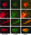
FIG. 1.
FIG. 1.
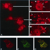
Indirect immunofluorescence analysis of VP40 and NP distribution in MBGV-infected human macrophages. Human macrophages were infected with MBGV and fixed at 48 h p.i. Immunofluorescence analysis was performed with an anti-VP40 monoclonal antibody (dilution, 1:100) and a secondary donkey anti-mouse antibody conjugated with rhodamine (dilution, 1:100), together with a guinea pig anti-NP serum (dilution, 1:100) and a secondary donkey anti-guinea pig antibody conjugated with FITC (dilution, 1:100). (A, D, G, and J) Anti-VP40/rhodamine. (B, E, H, and K) Anti-NP/FITC. (C, F, I, and L) Merged images. The arrowheads indicate colocalization of VP40 and NP. The arrows indicate singly located VP40.
FIG. 2.
FIG. 2.
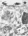
Immunoelectron microscopic analysis of VP40 and NP distribution in MBGV-infected human macrophages. Human macrophages were infected with MBGV and fixed at 48 h p.i. (A to D) Immunoelectron microscopic analysis was performed with an anti-VP40 monoclonal antibody (dilution, 1:100) and a secondary donkey anti-mouse antibody conjugated with colloidal gold (bead diameter, 6 nm; dilution, 1:25), together with a guinea pig anti-NP serum (dilution, 1:100) and a secondary donkey anti-guinea pig antibody conjugated with colloidal gold (bead diameter, 12 nm; dilution, 1:25). The black and white arrows indicate VP40 (6-nm gold beads). The arrowheads indicate sites of VP40-positive double membranes. Bar, 100 nm. (A) Focus of viral budding. (B) Multivesicular body in direct vicinity of a viral inclusion. (C) Multivesicular body in cytoplasm. (D) Unfolded multivesicular body beneath plasma membrane. (E) Transmission electron microscopy. The arrow indicates viral particles. The arrowheads indicate sites of double membranes. Vi, viral inclusion. Bar, 1,000 nm. (F) Presence of VP40 in protrusions (12-nm gold beads). Bar, 100 nm. (G) NP inside the protrusions is restricted to virions (6-nm gold bead). Bar, 100 nm.
FIG. 2.
FIG. 2.
Immunoelectron microscopic analysis of VP40 and NP distribution in MBGV-infected human macrophages. Human macrophages were infected with MBGV and fixed at 48 h p.i. (A to D) Immunoelectron microscopic analysis was performed with an anti-VP40 monoclonal antibody (dilution, 1:100) and a secondary donkey anti-mouse antibody conjugated with colloidal gold (bead diameter, 6 nm; dilution, 1:25), together with a guinea pig anti-NP serum (dilution, 1:100) and a secondary donkey anti-guinea pig antibody conjugated with colloidal gold (bead diameter, 12 nm; dilution, 1:25). The black and white arrows indicate VP40 (6-nm gold beads). The arrowheads indicate sites of VP40-positive double membranes. Bar, 100 nm. (A) Focus of viral budding. (B) Multivesicular body in direct vicinity of a viral inclusion. (C) Multivesicular body in cytoplasm. (D) Unfolded multivesicular body beneath plasma membrane. (E) Transmission electron microscopy. The arrow indicates viral particles. The arrowheads indicate sites of double membranes. Vi, viral inclusion. Bar, 1,000 nm. (F) Presence of VP40 in protrusions (12-nm gold beads). Bar, 100 nm. (G) NP inside the protrusions is restricted to virions (6-nm gold bead). Bar, 100 nm.
FIG. 3.
FIG. 3.
Indirect immunofluorescence analysis of recombinant VP40 in HeLa cells. HeLa cells were infected with MVA-T7 and transfected with pTM-VP40. At 16 h posttransfection, cells were fixed and immunostained with an anti-VP40 monoclonal antibody (dilution, 1:100) and a secondary donkey anti-mouse IgG conjugated with rhodamine (dilution, 1:100). (A) Distribution of recombinant VP40. The framed parts are shown at a higher magnification in panels B to D. The arrows indicate the tubular network; the arrowheads indicate bright spots. (E to G) Colocalization of actin and VP40 (confocal micrograph). (E) Distribution of VP40. (F) Distribution of filamentous actin. (G) Merge of panels E and F.
FIG. 4.
FIG. 4.
Membrane association of VP40. (A) HeLa cells were infected with MVA-T7 and transfected with pTM-40. The cells were harvested and lysed at 6, 12, and 16 h posttransfection. Sucrose was added to the postnuclear supernatant to a final concentration of 63%. The sample was placed at the bottom of centrifuge tube and overlaid with 45 and 10% sucrose. The gradient was centrifuged to equilibrium at 35,000 rpm overnight in an SW41 rotor. Fractions were collected from the top, and samples were separated by SDS-PAGE and blotted onto polyvinylidene difluoride membranes. Membranes were stained with an anti-VP40 monoclonal antibody (dilution, 1:1,000). Top of the gradient, fraction 1; bottom of the gradient, fraction 11. (B) Triton X-114 phase-partitioning analysis of VP40 at 16 h posttransfection. HeLa cells were infected and transfected as described for panel A. The postnuclear supernatant was partitioned into aqueous (lane A), detergent (lane D), and insoluble (lane P) fractions as described in Materials and Methods. Staining was done with an anti-LAMP-1 monoclonal antibody (dilution, 1:250) and with an anti-VP40 monoclonal antibody (dilution, 1:1,000). (C) Characterization of the membrane association of VP40 at 16 h posttransfection. Flotation analysis of postnuclear supernatant of recombinant VP40 was carried out as described for panel A. The postnuclear supernatant was treated before flotation analysis with either 2 M KCl (upper panel), EDTA (middle panel), or high pH (lower panel). Western blots were stained with an anti-VP40 monoclonal antibody (dilution, 1:1,000). (D) Characterization of cellular proteins comigrating with VP40 in flotation analysis. Western blots were stained with monoclonal antibodies directed against marker proteins of plasma membrane (VLA-2α), late endosomes (Lamp-1), endoplasmic reticulum (BiP/GRP78), and annexin II.
FIG. 5.
FIG. 5.
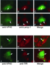
Immunofluorescence analysis of the intracellular distribution of VP40, Lamp-1, and TfR. HeLa cells were infected with MVA-T7 and transfected with pTM-VP40. At 16 h posttransfection, cells were fixed and immunostained with a rabbit anti-VP40 antibody (dilution, 1:10) and a secondary donkey anti-rabbit IgG conjugated with FITC (dilution, 1:100), together with either an anti-Lamp-1 (A to F) or an anti-TfR (G to L) monoclonal antibody (dilution, 1:20) and a secondary donkey anti-mouse antibody conjugated with rhodamine. (A and D) VP40/FITC. (B and E) Lamp-1-rhodamine. (C and F) Merged images. The arrows indicate the colocalization of VP40 and Lamp-1 in the tubular network (A to C) or in spots (D to F). (E and F) The arrowheads indicate a Lamp-1-positive spot in a VP40-negative cell. (G and J) VP40-FITC. (H and K) TfR-rhodamine. (C and F) Merged images. The arrows indicate sites of overlapping in leading edge of the cell (G to I) or in spots (J to L). (K and L) The arrowheads indicate a TfR-positive leading edge that does not contain VP40.
FIG. 6.
FIG. 6.
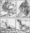
Immunoelectron microscopic analysis of recombinant VP40 in HeLa cells. HeLa cells were infected with MVA-T7 and transfected with pTM-VP40. At 16 h posttransfection, cells were fixed and immunostained with either a rabbit affinity-purified anti-VP40 antiserum (dilution, 1:10) and an anti-Lamp-1 monoclonal antibody (dilution, 1:20) (A, B, and D) or an anti-VP40 monoclonal antibody (dilution, 1:100) and rabbit IgG against actin (dilution, 1:200) (C). Donkey anti-mouse antibody conjugated with colloidal gold (bead diameter, 6 nm) and donkey anti-rabbit antibody conjugated with colloidal gold (bead diameter, 12 nm) were used as secondary antibodies. (A, B, and D) The arrows indicate Lamp-1 (6-nm gold beads). N, nucleus. In panel D, the arrowhead indicates a site of double membranes. (C) The arrows indicate VP40 (6-nm gold beads), and the arrowheads indicate actin (12-nm gold beads). Bar, 100 nm.
FIG. 7.
FIG. 7.
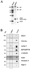
Western blot analysis of cellular proteins in purified MBGV structural proteins. Purified MBGV structural proteins (20) were separated by SDS-PAGE and either stained with Coomassie blue or blotted onto polyvinylidene difluoride membranes. (A) Coomassie blue staining of purified MBGV structural proteins. (B) Western blot analysis of cellular proteins in purified virions. Mouse monoclonal antibodies directed against marker proteins of early endosomes (EEA1), plasma membrane (VLA-2α), late endosomes (Lamp-1), endoplasmic reticulum (BiP/GRP78), microtubules (α-tubulin), annexin II, and small GTPase (Rab11), or rabbit IgG directed against actin were used to detect the respective proteins. Control, uninfected HeLa cells.
FIG. 8.
FIG. 8.
Immunoelectron microscopic analysis of detergent-treated MBGV particles. Purified virions were treated with Triton X-100 (A and B) or left untreated as a control (C) and immunostained with an anti-VP40 mouse monoclonal antibody and a secondary donkey anti-mouse antibody conjugated with colloidal gold (6-nm gold beads, arrows). White lines indicate regular striation of the viral envelop. Bar, 100 nm.

Similar articles

Cited by

References

    1. Becker, S., H.-D. Klenk, and E. Mühlberger. 1996. Intracellular transport and processing of the Marburg virus surface protein in vertebrate and insect cells. Virology 225:145-155. - PubMed
    1. Becker, S., C. Rinne, U. Hofsäss, H.-D. Klenk, and E. Mühlberger. 1998. Interactions of Marburg virus nucleocapsid proteins. Virology 249:406-417. - PubMed
    1. Bergmann, J. E., and P. J. Fusco. 1988. The M protein of vesicular stomatitis virus associates specifically with the basolateral membranes of polarized epithelial cells independently of the G protein. J. Cell Biol. 107:1707-1715. - PMC - PubMed
    1. Bordier, C. 1981. Phase separation of integral membrane proteins in Triton X-114 solution. J. Biol. Chem. 256:1604-1607. - PubMed
    1. Bretscher, M. S., and C. Aguado-Velasco. 1998. Membrane traffic during cell locomotion. Curr. Opin. Cell Biol. 10:537-541. - PubMed

Publication types

LinkOut - more resources