

Internalization of echovirus 1 in caveolae
- PMID: 11799180
- PMCID: PMC135881
- DOI: 10.1128/jvi.76.4.1856-1865.2002
Internalization of echovirus 1 in caveolae
Abstract
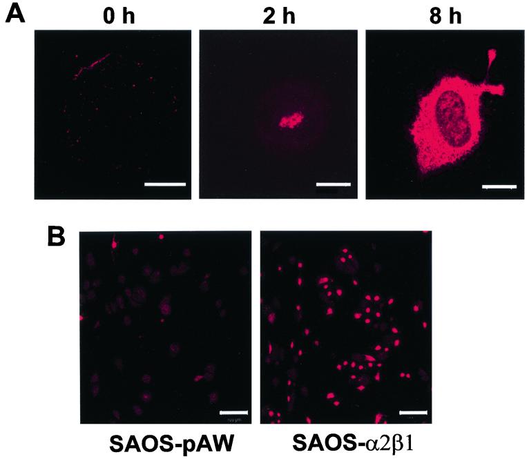
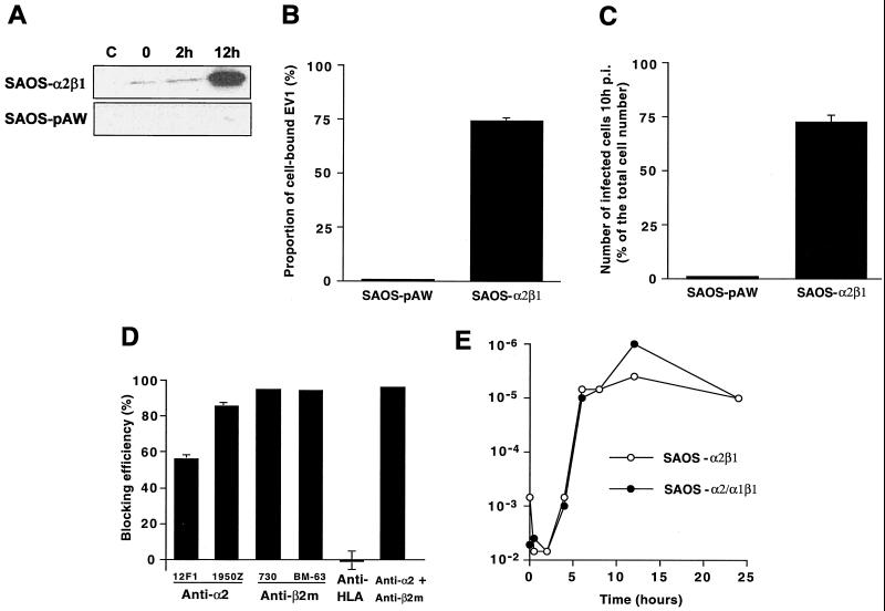
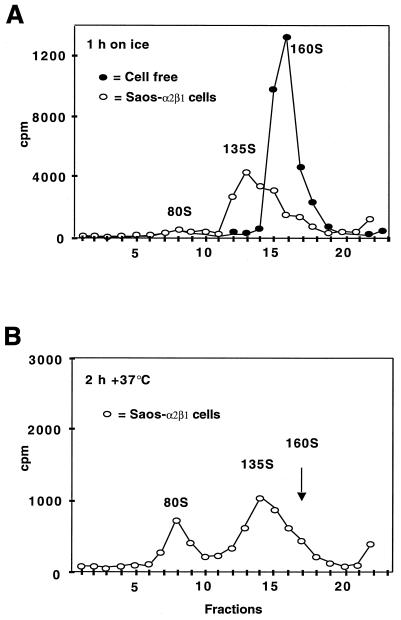
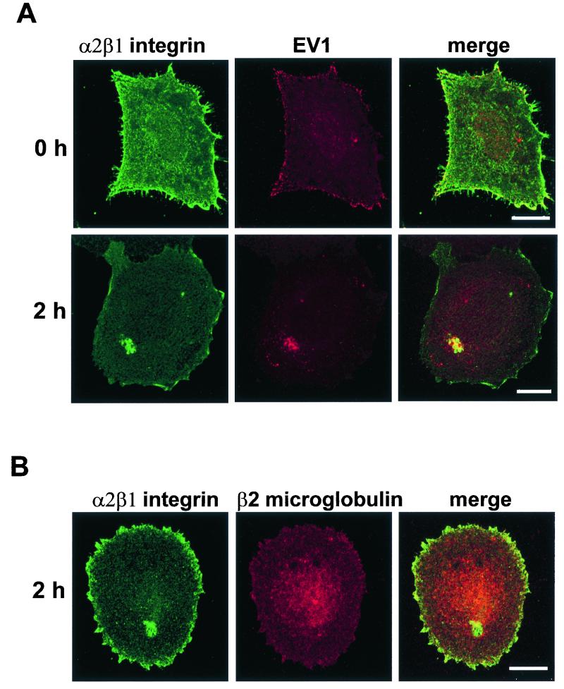
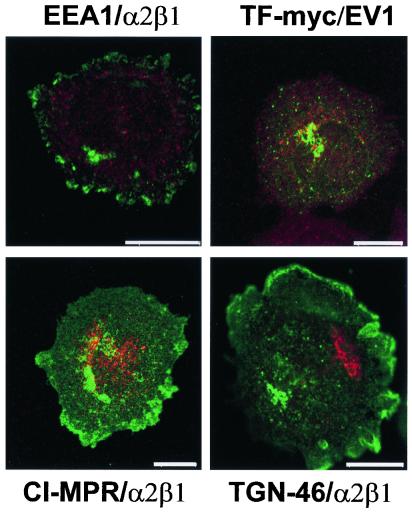
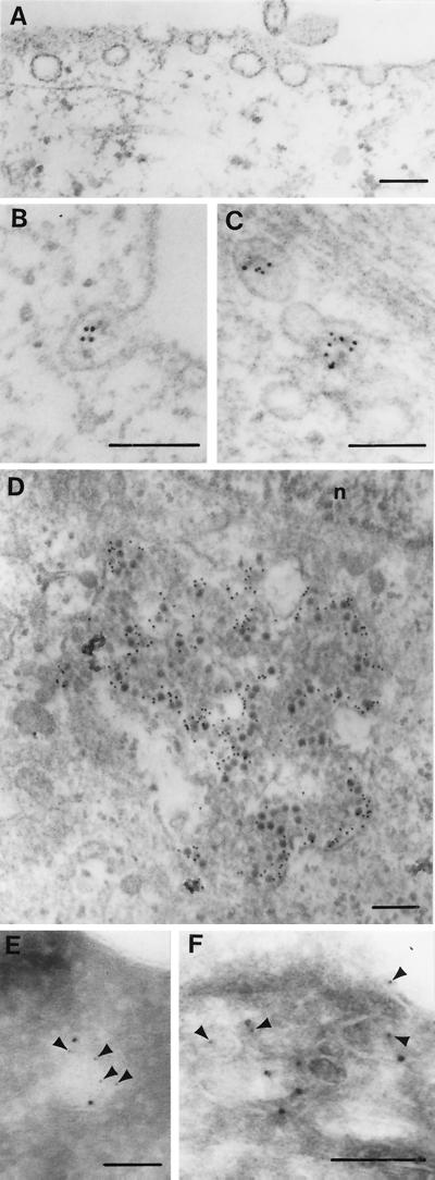
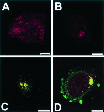
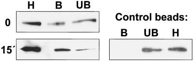
Echovirus 1 (EV1) is a human pathogen which belongs to the Picornaviridae family of RNA viruses. We have analyzed the early events of infection after EV1 binding to its receptor alpha 2 beta 1 integrin and elucidated the route by which EV1 gains access to the host cell. EV1 binding onto the cell surface and subsequent entry resulted in conformational changes of the viral capsid as demonstrated by sucrose gradient sedimentation analysis. After 15 min to 2 h postinfection (p.i.) EV1 capsid proteins were seen in vesicular structures that were negative for markers of the clathrin-dependent endocytic pathway. In contrast, immunofluorescence confocal microscopy showed that EV1, alpha 2 beta 1 integrin, and caveolin-1 were internalized together in vesicular structures to the perinuclear area. Electron microscopy showed the presence of EV1 particles inside caveolae. Furthermore, infective EV1 could be isolated with anti-caveolin-1 beads 15 min p.i., confirming a close association with caveolin-1. Finally, the expression of dominant negative caveolin in cells markedly inhibited EV1 infection, indicating the importance of caveolae for the viral replication cycle of EV1.
Figures
Similar articles
-
Clustering induces a lateral redistribution of alpha 2 beta 1 integrin from membrane rafts to caveolae and subsequent protein kinase C-dependent internalization.Mol Biol Cell. 2004 Feb;15(2):625-36. doi: 10.1091/mbc.e03-08-0588. Epub 2003 Dec 2. Mol Biol Cell. 2004. PMID: 14657242 Free PMC article.
-
Structural and functional analysis of integrin alpha2I domain interaction with echovirus 1.J Biol Chem. 2004 Mar 19;279(12):11632-8. doi: 10.1074/jbc.M312441200. Epub 2003 Dec 29. J Biol Chem. 2004. PMID: 14701832
-
Echovirus 1 endocytosis into caveosomes requires lipid rafts, dynamin II, and signaling events.Mol Biol Cell. 2004 Nov;15(11):4911-25. doi: 10.1091/mbc.e04-01-0070. Epub 2004 Sep 8. Mol Biol Cell. 2004. PMID: 15356270 Free PMC article.
-
Caveolae and caveolin isoforms in rat peritoneal macrophages.Micron. 2002;33(1):75-93. doi: 10.1016/s0968-4328(00)00100-1. Micron. 2002. PMID: 11473817 Review.
-
Caveolae/raft-dependent endocytosis.J Cell Biol. 2003 May 26;161(4):673-7. doi: 10.1083/jcb.200302028. J Cell Biol. 2003. PMID: 12771123 Free PMC article. Review.
Cited by
-
Tailoring Iron Oxide Nanoparticles for Efficient Cellular Internalization and Endosomal Escape.Nanomaterials (Basel). 2020 Sep 11;10(9):1816. doi: 10.3390/nano10091816. Nanomaterials (Basel). 2020. PMID: 32932957 Free PMC article. Review.
-
Function of membrane rafts in viral lifecycles and host cellular response.Biochem Res Int. 2011;2011:245090. doi: 10.1155/2011/245090. Epub 2011 Dec 7. Biochem Res Int. 2011. PMID: 22191032 Free PMC article.
-
Pseudomonas aeruginosa-mediated damage requires distinct receptors at the apical and basolateral surfaces of the polarized epithelium.Infect Immun. 2010 Mar;78(3):939-53. doi: 10.1128/IAI.01215-09. Epub 2009 Dec 14. Infect Immun. 2010. PMID: 20008530 Free PMC article.
-
Localization of Ptr ToxA Produced by Pyrenophora tritici-repentis Reveals Protein Import into Wheat Mesophyll Cells.Plant Cell. 2005 Nov;17(11):3203-12. doi: 10.1105/tpc.105.035063. Epub 2005 Sep 30. Plant Cell. 2005. PMID: 16199615 Free PMC article.
-
Monkey rotavirus binding to alpha2beta1 integrin requires the alpha2 I domain and is facilitated by the homologous beta1 subunit.J Virol. 2003 Sep;77(17):9486-501. doi: 10.1128/jvi.77.17.9486-9501.2003. J Virol. 2003. PMID: 12915563 Free PMC article.
References
-
- Anderson, H. A., Y. Chen, and L. C. Norkin. 1998. MHC class I molecules are enriched in caveolae but do not enter with simian virus 40. J. Gen. Virol. 79:1469-1477. - PubMed
-
- Banting, G., R. Maile, and E. P. Roquemore. 1998. The steady state distribution of humTGN46 is not significantly altered in cells defective in clathrin-mediated endocytosis. J. Cell Sci. 111:3451-3458. - PubMed
-
- Barnstable, C. J., E. A. Jones, and M. J. Crumpton. 1978. Isolation, structure and genetics of HLA-A, -B, -C and -DRw (Ia) antigens. Br. Med. Bull. 34:241-246. - PubMed
Publication types
MeSH terms
Substances
LinkOut - more resources
Full Text Sources
Other Literature Sources
