. 2002 Feb;76(4):1884-91.
doi: 10.1128/jvi.76.4.1884-1891.2002.

The VP1 N-terminal sequence of canine parvovirus affects nuclear transport of capsids and efficient cell infection

Affiliations

The VP1 N-terminal sequence of canine parvovirus affects nuclear transport of capsids and efficient cell infection

Maija Vihinen-Ranta et al. J Virol. 2002 Feb.

Abstract

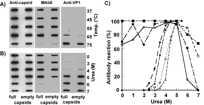
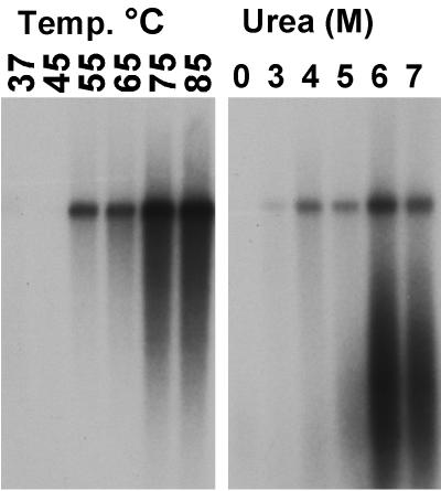
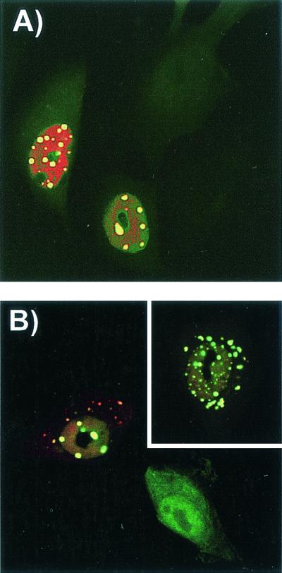
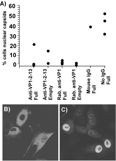
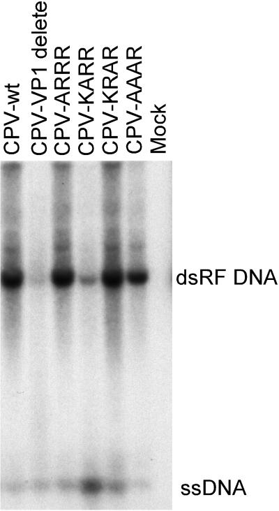
The unique N-terminal region of the parvovirus VP1 capsid protein is required for infectivity by the capsids but is not required for capsid assembly. The VP1 N terminus contains a number of groups of basic amino acids which resemble classical nuclear localization sequences, including a conserved sequence near the N terminus comprised of four basic amino acids, which in a peptide can act to transport other proteins into the cell nucleus. Testing with a monoclonal antibody recognizing residues 2 to 13 of VP1 (anti-VP1-2-13) and with a rabbit polyclonal serum against the entire VP1 unique region showed that the VP1 unique region was not exposed on purified capsids but that it became exposed after treatment of the capsids with heat (55 to 75 degrees C), or urea (3 to 5 M). A high concentration of anti-VP1-2-13 neutralized canine parvovirus (CPV) when it was incubated with the virus prior to inoculation of cells. Both antibodies blocked infection when injected into cells prior to virus inoculation, but neither prevented infection by coinjected infectious plasmid DNA. The VP1 unique region could be detected 4 and 8 h after the virus capsids were injected into cells, and that sequence exposure appeared to be correlated with nuclear transport of the capsids. To examine the role of the VP1 N terminus in infection, we altered that sequence in CPV, and some of those changes made the capsids inefficient at cell infection.


Figures

FIG. 1.
FIG. 1.
Exposure of the VP1 unique region detected by antibodies after urea or temperature treatments of full or empty capsids. The VP1 sequence was detected with rabbit anti-VP1 or with anti-VP1-2-13 MAb, intact capsids were detected with MAb 8, and the total capsid proteins were detected with a rabbit anticapsid antibody. (A) Treatment of capsids by heating for 30 min at the indicated temperatures prior to applying to the membrane. (B) Treatment of capsids with various urea concentrations for 30 min prior to applying to the membrane. (C) Antibody binding to full or empty capsids after treatment with various concentrations of urea. Data represents the means from four separate experiments and are presented as percentages of the maximal binding seen for each antibody. Open symbols, empty capsids; closed symbols, full capsids; circles, MAb 8; squares, rabbit anticapsid; diamonds, anti-VP1-2-13 MAb; triangles, rabbit anti-VP1.
FIG. 2.
FIG. 2.
Exposure of the 3" end of the viral DNA after temperature or heat treatment of full capsids. After treatments, capsids were washed and incubated with T7 DNA polymerase and 35S-dATP, and then the products were electrophoresed in agarose gels and detected on X-ray film. The labeled DNA band is the monomer-length double-stranded form produced from the viral genome.
FIG. 3.
FIG. 3.
Exposure of the VP1 unique region on purified capsids injected into cells. Full virus capsids were injected into cells, which were then incubated for 1, 4, or 8 h at 37°C before fixation. The cells were incubated with rabbit anti-VP1 unique region followed by TxR-conjugated anti-rabbit IgG. Capsids were detected using MAb 8 and FITC-conjugated anti-mouse IgG. Confocal images show a 1-μm section from the center of each cell, while phase-contrast images of the same cells are also shown.
FIG. 4.
FIG. 4.
Effect of injection with anti-VP1-2-13 or rabbit IgG against the complete VP1 unique region on infection of the cells by CPV. (A and B) Injection of anti-VP1-2-13 antibody (A) or control mouse IgG (B). The cells were costained for IgG (FITC, green) and for viral NS1 protein (red). (C) Percentage of cells that became infected after injection with anti-VP1-2-13, with the rabbit anti-VP1 unique region, or with control IgG or without injection in the same cultures. Error bars indicate standard deviations.
FIG. 5.
FIG. 5.
Detection of CPV replication and of capsid protein localization in cells injected simultaneously with anti-VP1-2-13 and the CPV infectious plasmid and then incubated for 24 h at 37°C. (A) Expression of NS1 in cells coinjected with anti-VP1-2-13 and a CPV infectious plasmid. The anti-VP1-2-13 was detected with an FITC-labeled anti-mouse IgG (green), and NS1 was detected with a TxR-labeled anti-NS1 IgG (red). (B) Localization of CPV capsid proteins in cells coinjected with anti-VP1-2-13 and a CPV infectious plasmid. The injected MAb was detected with FITC-labeled anti-mouse IgG (green), and capsid proteins were detected with a specific rabbit anticapsid IgG followed by TxR-labeled anti-rabbit IgG (red). The inset shows another cell subjected to the same treatment.
FIG. 6.
FIG. 6.
Nuclear localization of full or empty CPV capsids after injection into the cytoplasm of cells in the presence or absence of anti-VP1-2-13, rabbit (Rab.) anti-VP1, or control mouse IgG. The cells were incubated for 6 h after injection and then fixed and stained for capsids with a directly conjugated anticapsid antibody. (A) Percentage of cells injected with capsids showing substantial nuclear localization after 6 h of incubation. The data for each experiment are shown for each capsid and antibody combination. (B and C) Representative fields of cells showing the localization of capsids within cells 6 h after injection of virus and antibody (B) or virus alone (C) into the cells.
FIG. 7.
FIG. 7.
Production of replicative-form DNA (dsRF DNA) and genomic ssDNA after transfection of cells with the wild-type (CPV-wt) or mutant (Table 1) infectious plasmids. The DNA recovered by a modified Hirt lysis procedure was digested with DpnI prior to electrophoresis in a 1% agarose gel with 1 μg of ethidium bromide per ml. The viral DNAs were detected by Southern blotting.
FIG. 8.
FIG. 8.
Alignment of the VP1 N-terminal sequences from viruses related to CPV. More distantly related parvoviruses showed no significant homology in this region of the protein.

Similar articles

Cited by

References

    1. Agbandje, M., R. McKenna, M. G. Rossmann, M. L. Strassheim, and C. R. Parrish. 1993. Structure determination of feline panleukopenia virus empty particles. Proteins 16:155-171. - PubMed
    1. Anderson, S., M. Momoeda, M. Kawase, S. Kajigaya, and N. S. Young. 1995. Peptides derived from the unique region of B19 parvovirus minor capsid protein elicit neutralizing antibodies in rabbits. Virology 206:626-632. - PubMed
    1. Bui, M., G. Whittaker, and A. Helenius. 1996. Effect of M1 protein and low pH on nuclear transport of influenza virus ribonucleoproteins. J. Virol. 70:8391-8401. - PMC - PubMed
    1. Casal, J. I., J. P. Langeveld, E. Cortes, W. W. Schaaper, E. van Dijk, C. Vela, S. Kamstrup, and R. H. Meloen. 1995. Peptide vaccine against canine parvovirus: identification of two neutralization subsites in the N terminus of VP2 and optimization of the amino acid sequence. J. Virol. 69:7274-7277. - PMC - PubMed
    1. Chang, S. F., J. Y. Sgro, and C. R. Parrish. 1992. Multiple amino acids in the capsid structure of canine parvovirus coordinately determine the canine host range and specific antigenic and hemagglutination properties. J. Virol. 66:6858-6867. - PMC - PubMed

Publication types

MeSH terms

LinkOut - more resources