Regulated gene expression in the chicken embryo by using replication-competent retroviral vectors
- PMID: 11799192
- PMCID: PMC135918
- DOI: 10.1128/jvi.76.4.1980-1985.2002
Regulated gene expression in the chicken embryo by using replication-competent retroviral vectors
Abstract
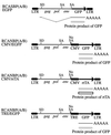
Rous sarcoma virus (RSV)-derived retroviral vector could efficiently deliver the green fluorescent protein (GFP), which is driven by the internal cytomegalovirus enhancer/promoter, into restricted cell populations in the chicken embryo. RSV-derived vectors coupled with the tet regulatory elements also revealed doxycycline-dependent inducible GFP expression in the chicken embryo in ovo.
Figures


Similar articles
-
Gene delivery into the chicken embryo by using replication-competent retroviral vectors.Fukushima J Med Sci. 2004 Dec;50(2):37-46. doi: 10.5387/fms.50.37. Fukushima J Med Sci. 2004. PMID: 15779569 Review.
-
Expression of Rous sarcoma virus-derived retroviral vectors in the avian blastoderm: potential as stable genetic markers.Proc Natl Acad Sci U S A. 1991 Dec 1;88(23):10505-9. doi: 10.1073/pnas.88.23.10505. Proc Natl Acad Sci U S A. 1991. PMID: 1660139 Free PMC article.
-
Replication-competent retroviral vectors for expressing genes in avian cells in vitro and in vivo.Mol Biotechnol. 1997 Jun;7(3):289-98. doi: 10.1007/BF02740819. Mol Biotechnol. 1997. PMID: 9219242 Review.
-
Inducible retroviral vectors regulated by lac repressor in mammalian cells.Gene. 1996 Dec 12;183(1-2):137-42. doi: 10.1016/s0378-1119(96)00532-x. Gene. 1996. PMID: 8996098
-
Gene transfer into mammalian cells by a Rous sarcoma virus-based retroviral vector with the host range of the amphotropic murine leukemia virus.J Virol. 1996 Jun;70(6):3922-9. doi: 10.1128/JVI.70.6.3922-3929.1996. J Virol. 1996. PMID: 8648729 Free PMC article.
Cited by
-
The function of neurotrophic factor receptors expressed by the developing adductor motor pool in vivo.J Neurosci. 2004 May 12;24(19):4668-82. doi: 10.1523/JNEUROSCI.0580-04.2004. J Neurosci. 2004. PMID: 15140938 Free PMC article.
-
Small GTPase protein Rac-1 is activated with maturation and regulates cell morphology and function in chondrocytes.Exp Cell Res. 2008 Apr 1;314(6):1301-12. doi: 10.1016/j.yexcr.2007.12.029. Epub 2008 Jan 18. Exp Cell Res. 2008. PMID: 18261726 Free PMC article.
-
A Tet/Q Hybrid System for Robust and Versatile Control of Transgene Expression in C. elegans.iScience. 2019 Jan 25;11:224-237. doi: 10.1016/j.isci.2018.12.023. Epub 2018 Dec 27. iScience. 2019. PMID: 30634168 Free PMC article.
-
Study of the regulatory elements of the Ovalbumin gene promoter using CRISPR technology in chicken cells.J Biol Eng. 2023 Jul 17;17(1):46. doi: 10.1186/s13036-023-00367-3. J Biol Eng. 2023. PMID: 37461059 Free PMC article.
-
Ex vivo electroporation of retinal cells: a novel, high efficiency method for functional studies in primary retinal cultures.Exp Eye Res. 2013 Apr;109:40-50. doi: 10.1016/j.exer.2013.01.010. Epub 2013 Jan 28. Exp Eye Res. 2013. PMID: 23370269 Free PMC article.
References
-
- Baskar, J. F., P. P. Smith, G. Nilaver, R. A. Jupp, S. Hoffmann, N. J. Peffer, D. J. Tenney, A. M. Colberg-Poley, P. Ghazal, and J. A. Nelson. 1996. The enhancer domain of the human cytomegalovirus major immediate early promoter determines cell type-specific expression in transgenic mice. J. Viol. 70:3207-3214. - PMC - PubMed
-
- Bello, B., D. Resendez-Perez, and W. J. Gehring. 1998. Spatial and temporal targeting of gene expression in Drosophila by means of a tetracycline-dependent transactivator system. Development 125:2193-2202. - PubMed
-
- Blaauw, M., M. H. Linskens, and P. J. van Haastert. 2000. Efficient control of gene expression by a tetracycline-dependent transactivator in single Dictyostelium discoideum cells. Gene 252:71-82. - PubMed
-
- Briscoe, J., A. Pierani, T. M. Jessell, and J. Ericson. 2000. A homeodomain protein code specifies progenitor cell identity and neuronal fate in the ventral neural tube. Cell 101:435-445. - PubMed
-
- Federspiel, M. J., and S. H. Hughes. 1997. Retroviral gene delivery. Methods Cell Biol. 52:179-214. - PubMed
Publication types
MeSH terms
Substances
LinkOut - more resources
Full Text Sources

