. 2002 Jan;86(1):62-9.
doi: 10.1136/bjo.86.1.62.

Clinicopathological correlation of congenital corneal opacification using ultrasound biomicroscopy

Affiliations

Clinicopathological correlation of congenital corneal opacification using ultrasound biomicroscopy

K K Nischal et al. Br J Ophthalmol. 2002 Jan.

Abstract

Aim: To investigate the correlation between clinical, high frequency ultrasound biomicroscopy (UBM) and, where possible, histological findings in cases of congenital corneal opacification presenting to the departments of ophthalmology, Great Ormond Street Hospital for Children, London, and the Hospital for Sick Children, Toronto, Canada.

Method: 22 eyes of 13 children (age range 3-225 days) with congenitally opaque corneas were examined. UBM was performed using the ultrasound biomicroscope (Allergan-Humphrey). All eyes underwent penetrating keratoplasties (PKP) except five. The host corneas were all sent for histological examination.

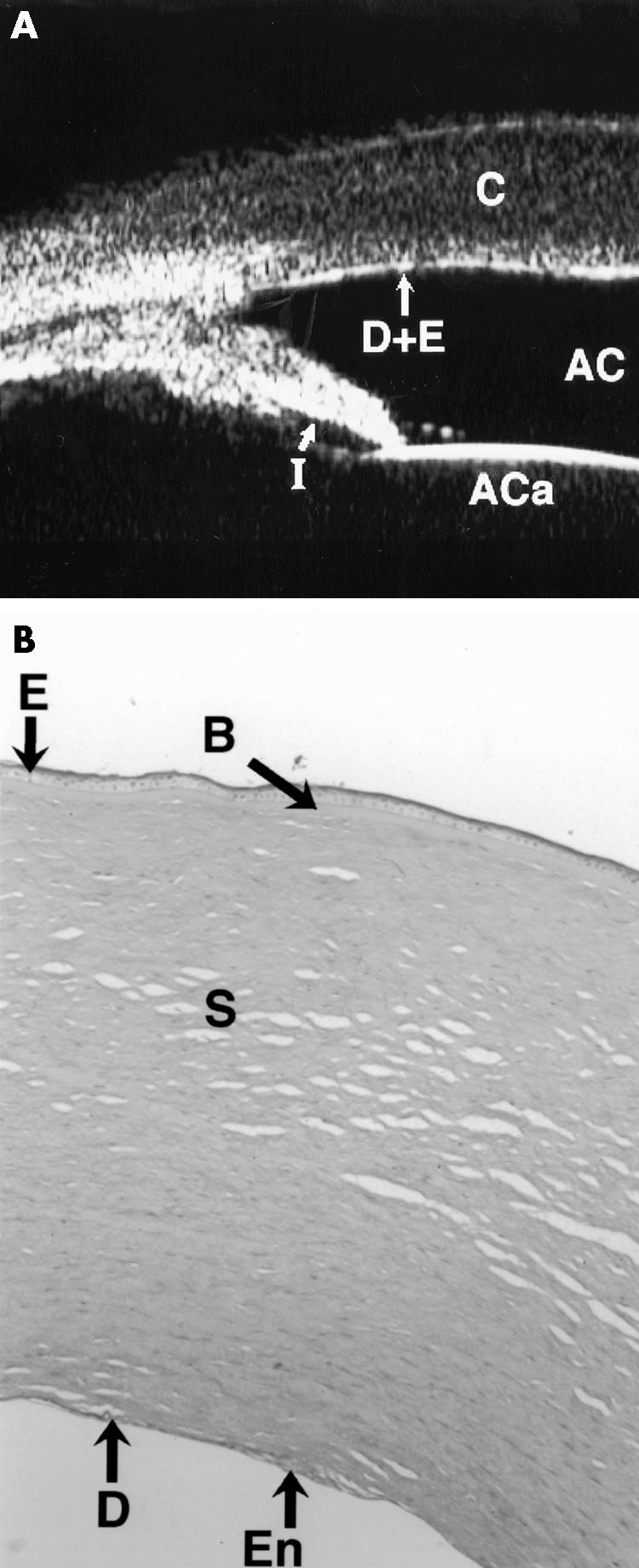
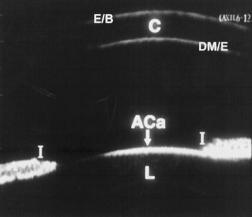
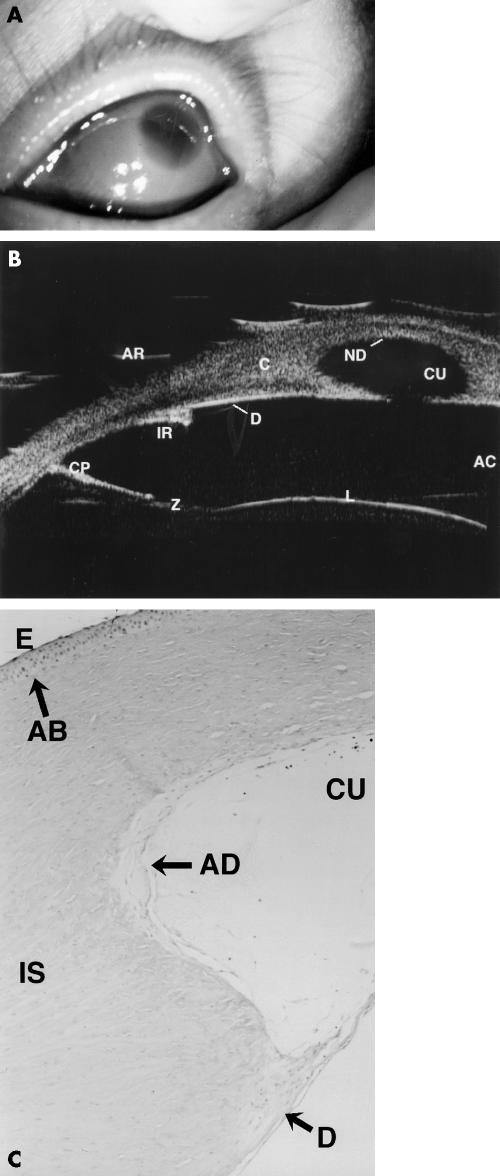
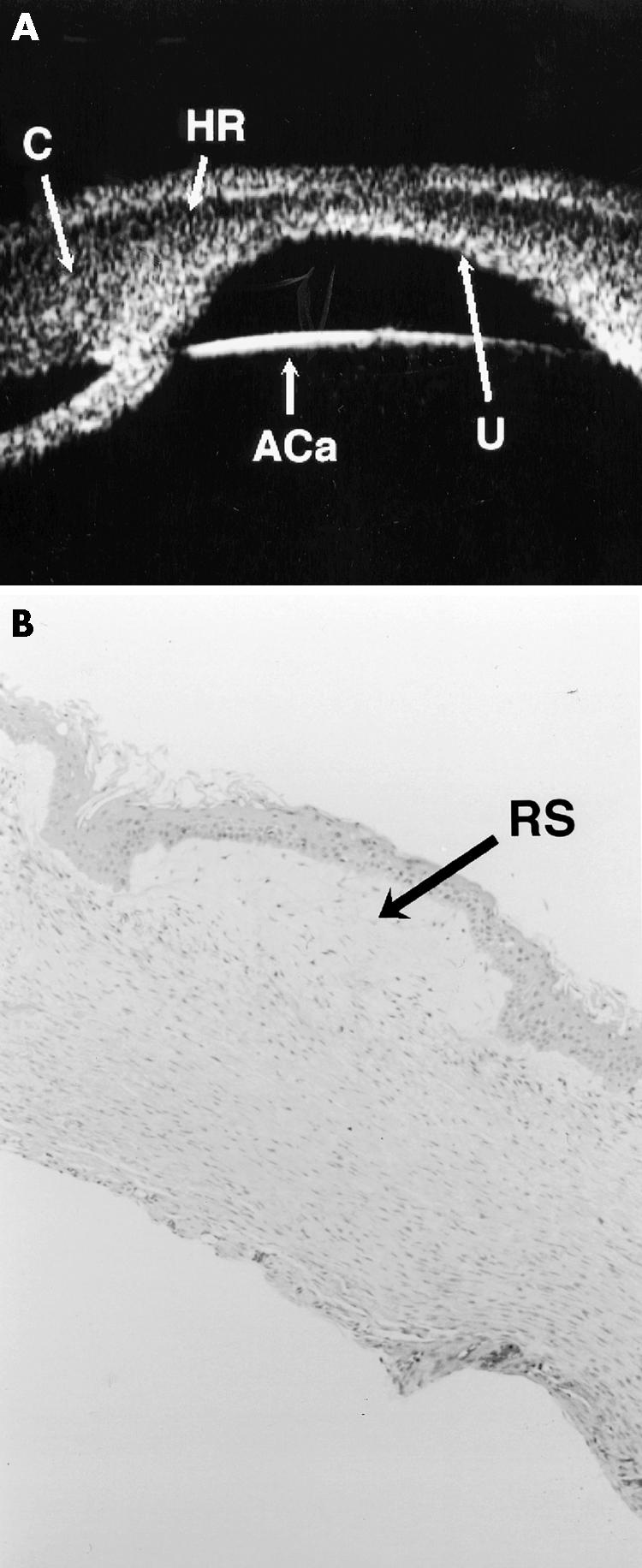
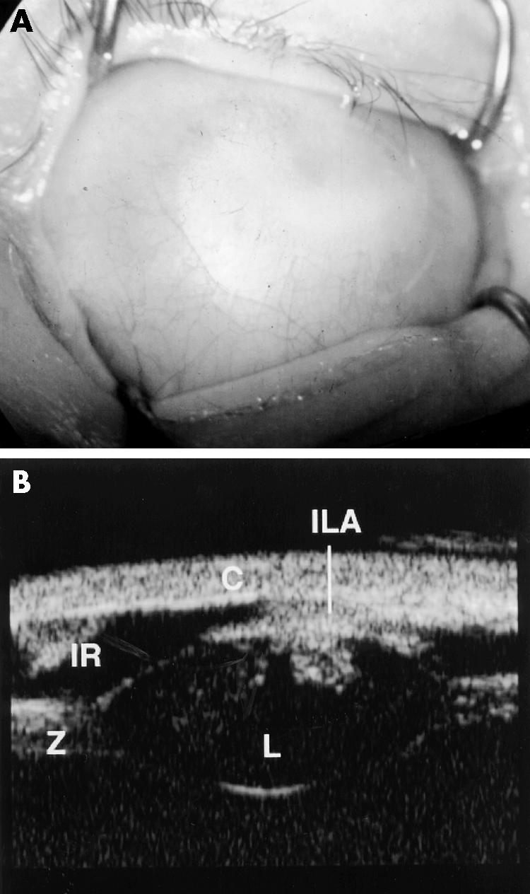
Results: The final diagnosis in our series was Peters' anomaly in nine cases (70%), corneal dystrophy in two cases (15%), and sclerocornea in two cases (15%). The UBM findings changed the clinical diagnosis in five cases (38%). In these five cases histology was available in four and confirmed the UBM diagnosis in each case. In no case of the 13 where histology was available did it contradict the UBM findings. In two cases a hypoechoic region in the anterior stroma was seen on UBM which correlated histologically with absent Bowman's layer and oedema. In two cases UBM revealed aniridia and in one, congenital aphakia, which was not apparent clinically.

Conclusion: UBM examination is not only very useful in evaluating the clinical diagnosis in congenital corneal opacification, it also acts as a preoperative guide in cases undergoing PKP by detecting keratolenticular and iridocorneal adhesions and other ocular abnormalities such as aniridia and congenital aphakia. In all cases where PKP was performed the UBM diagnosis was confirmed histologically. The clinical diagnosis was incorrect in five cases. This has important implications in studies of phenotype/genotype correlation of congenital corneal opacification.


Figures

Figure 1
Figure 1
(A) The UBM of case 1 shows irregular thickness of the cornea (C) and focal anomalies in the Descemet's membrane and endothelial echo (D+E) (compare with Fig 2). The pupil is dilated, and so the iris appears shorter than normal (I). The anterior chamber (AC) and the anterior capsule of the lens are also seen. The corneal thickness for the right eye was 2.3 mm centrally and 2.2 mm for the left eye centrally. A diagnosis of corneal dystrophy was made, which was confirmed histologically as being posterior polymorphous dystrophy. (B) Full thickness (×10) cornea section is shown with periodic acid Schiff (PAS) stain. There is normal epithelium (E) and Bowman's layer (B) but the Descemet's membrane (DM) is abnormally thin with multilayering of the endothelium seen (En).
Figure 2
Figure 2
UBM of a normal age matched eye. The echogenicity from the epithelial/Bowman's layer complex (E/B), cornea (C), Descemet's membrane/endothelial complex (DM/E), anterior capsule of the lens (ACa), lens (L), and iris (I) is shown.
Figure 3
Figure 3
Clinical, UBM, and histological findings in case 5. (A) Shows the corneal opacity with relative clearing centrally. (B) A composite of two UBM scans showing aniridia with an iris stump (IR), stretched ciliary processes (CP), zonules (Z), the lens (L), intact Descemet's membrane/endothelial echo (D), central defect in posterior cornea (CU), and loss of the Descemet's membrane/endothelial echo within the defect (ND). AR = artefact. C = cornea. AC = anterior chamber. (C) (×10) Periodic acid Schiff (PAS) stain section of cornea is shown. This demonstrates the central defect (CU) with absent Descemet's membrane and endothelium (AD), but a thin Descemet's membrane is seen peripherally (D). Bowman's layer is absent (AB) and stromal lamellae are irregular (IS). E = epithelium. The relative clarity centrally seen in (A) is due to the gross central defect.
Figure 4
Figure 4
UBM scans and histology of case 4. (A) Shows the cornea (C), with a hypoechoic region (HR) in the anterior stroma with a central defect in the posterior aspect of the cornea (U). The anterior capsule of the lens is seen (ACa) as is a central iridocorneal adhesion. (B) Full thickness corneal section (haematoxylin and eosin stain) shows absence of Bowman's layer with marked rarefaction of the subepithelial stroma (RS), which corresponds to the hypoechoic layer seen on UBM. There is absence of Descemet's membrane and endothelium. All these histological features are consistent with a diagnosis of Peters' anomaly.
Figure 5
Figure 5
Clinical and UBM findings in case 2. (A) Shows complete corneal opacification thought clinically to be sclerocornea. (B) The UBM of the same case shows keratolenticular adhesion (ILA), aniridia with only an iris stump detected (IR), a small lens (L), and thickened looking zonules (Z). The cornea is also seen (C). The diagnosis post UBM was thought to be Peters' anomaly given the keratolenticular adhesion. Case 2 did not have penetrating keratoplasty performed.

References

    1. Pavlin CJ, Sherar MD, Foster S. Subsurface ultrasound microscopic imaging of the intact eye. Ophthalmology 1990;97:244–50. - PubMed
    1. Pavlin CJ, Harasiewicz K, Sherar MD, et al. Clinical use of ultrasound biomicroscopy. Ophthalmology 1991;98:287–95. - PubMed
    1. Pavlin CJ. Interpreting technology; practical application of ultrasound biomicroscopy. Canad J Ophthalmol 1995;30:225–9. - PubMed
    1. Bermejo E, Martinez-Frias ML. Congenital eye malformations: clinical epidemiological analysis of 1,124,654 consecutive births in Spain. Am J Med Genet 1998;75:497–504. - PubMed
    1. Avitabile T, Russo V, Ghirlanda R, et al. Corneal oedemas: diagnosis and surgical planning with ultrasound biomicroscopy. Ophthalmologica 1998;212(Suppl 1):13–16. - PubMed