. 2002 Jan;86(1):97-101.
doi: 10.1136/bjo.86.1.97.

Adenovirus mediated gene delivery of tissue inhibitor of metalloproteinases-3 induces death in retinal pigment epithelial cells

Affiliations

Adenovirus mediated gene delivery of tissue inhibitor of metalloproteinases-3 induces death in retinal pigment epithelial cells

Mohammed A Majid et al. Br J Ophthalmol. 2002 Jan.

Abstract

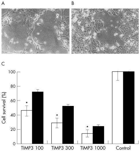
Background: Sorsby's fundus dystrophy (SFD) and age related macular degeneration (ARMD) are retinal diseases associated with a high level of accumulation of mutant and wild type TIMP-3, respectively, in Bruch's membrane. The pathogenic role of TIMP-3 in these diseases is uncertain, but causative mutations have been identified in the TIMP-3 gene of patients with SFD. Recent reports that TIMP-3 causes apoptosis in certain cell types and not in others prompted the authors to investigate whether TIMP-3 causes apoptosis in cultured retinal pigment epithelium (RPE) cells.

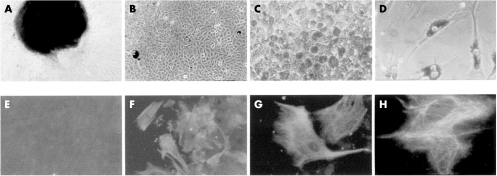
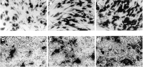
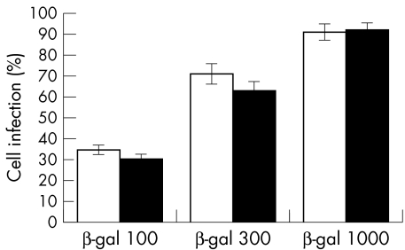
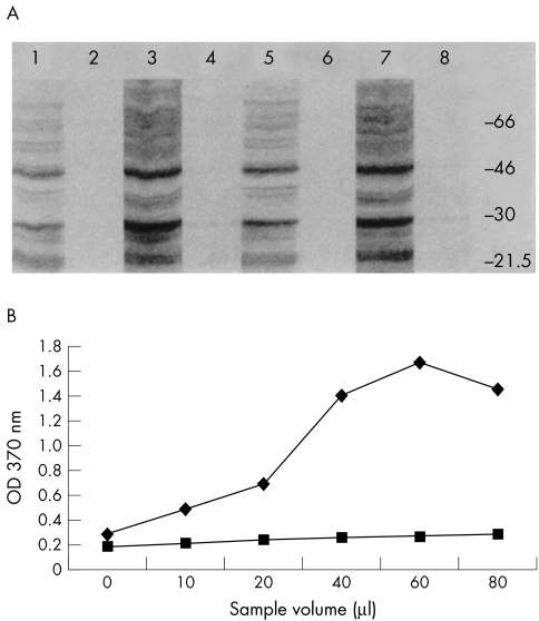
Methods: RPE and MCF-7 cells (as a positive control) were initially infected with replication deficient adenovirus, to overexpress beta-galactosidase (RAdLacZ) or TIMP-3 (RAdTIMP-3). TIMP-3 was detected by western blotting and ELISA. Cell viability was defined by cell counts. ISEL was used to investigate the mechanism of cell death.

Results: Cultured RPE cells produced small quantities of endogenous TIMP-3 and remained viable. However, overexpression of TIMP-3 caused a dose related death of RPE cells. The mechanism of cell death was apoptosis.

Conclusion: The previously unreported finding of TIMP-3 induced apoptosis of RPE cells may account for some of the early features seen in SFD and ARMD.


Figures

Figure 1
Figure 1
(A) Retinal explant giving rise to RPE cells (×100). (B) Confluent fusiform RPE cell culture lacking pigment (×100). (C) Pigmented polygonal RPE cell culture (×200). (D) Pigmented fusiform RPE cells (×200). (E) Corneal fibroblasts (negative control) immunostained for cytokeratins 8 and 18 (×200). (F, G, H): RPE cells immunostained for cytokeratins 8 and 18 (×200, ×400, and ×1000, respectively).
Figure 2
Figure 2
RAdlacZ infected RPE cells (A, B, C) and MCF7 cells (D, E, F) at 100, 300, and 1000 pfu/cell, respectively, stained with X-gal (×200).
Figure 3
Figure 3
A histogram of the numbers of X-gal positive RPE (open bars) and MCF-7 (solid bars) cells, 48 hours after infection with RadlacZ and staining for β-galactosidase activity. Statistical significance by Student's t test compared with corresponding group: p >0.2 in all groups (n=9).
Figure 4
Figure 4
(A) An immunostained western blot of the SDS extracted proteins from RPE and MCF7 cell matrices and 10-fold concentrated media fractions. Lane 1, RPE RAdTIMP-3 media; lane 2, RPE RAdlacZ media; lane 3, RPE RAdTIMP-3 matrix; lane 4, RPE RAdlacZ matrix; lane 5, MCF7 RAdTIMP-3 media; lane 6, MCF7 RAdlacZ media; lane 7, MCF7 RAdTIMP-3 matrix; lane 8, MCF7 RAdTIMP-3 media. (B) A line chart showing the results of ELISA detection of endogenous TIMP-3 in established RPE (upper line) and keratocyte (lower line) cell cultures. The control cells show relatively little endogenous TIMP-3 production compared with RPE cells.
Figure 5
Figure 5
(A, B) Representative photographs (×100) showing RPE (A) and MCF7 (B) cells 48 hours after infection with RAdTIMP-3. Note the cells rounding up and lifting, leaving patches of cell loss, consistent with localised TIMP-3 deposition. (C) A histogram of the numbers of viable RPE (open bars) and MCF7 (solid bars) cells remaining in culture 48 hours after infection with RAdTIMP-3. These cells were trypsinised, stained with trypan blue, and counted with a haemacytometer. Statistical significance, by Student's t test, compared with corresponding MCF7 group: *p <0.001 (n=9). Statistical significance, by Student's t test, compared with corresponding control group p<0.001 (n=9) for all groups.
Figure 6
Figure 6
Representative photographs (×100 magnification) of ISEL labelled, RAdTIMP-3 infected RPE and MCF7 cells (A and C, respectively) and RadlacZ infected cells (B and D, respectively). The apoptotic cells were detected with cobalt-DAB and are seen with darkly stained nuclei. The control cells were ISEL negative.

References

    1. Sorsby A, Mason MEJ. A fundus dystrophy with unusual features. Br J Ophthalmol 1949;33:67–97. - PMC - PubMed
    1. Capon MRC, Marshall J, Krafft JI, et al. Sorsby's fundus dystrophy. A light and electron microscopic study. Ophthalmology 1989;96:1769–77 - PubMed
    1. Polkinghorne PJ, Capon MRC, Berninger T, et al. Sorsby's fundus dystrophy. A clinical study. Ophthalmology 1989;96:1763–8. - PubMed
    1. Kamei M, Apte SS, Rayborn ME. TIMP-3 accumulation in Bruch's membrane and Drusen in eyes from normal and age-related macular degeneration donors. In: La Vail MM, Anderson RE, Hollyfield JG, eds. Degenerative diseases of the retina. New York: Plenum Press, 1997.
    1. Zarbin MA. Age-related macular degeneration: review of pathogenesis. Eur J Ophthalmol 1998;8:199–206. - PubMed

Publication types

MeSH terms

Substances