Comparative Study
. 2002 Jan 1;115(Pt 1):39-50.
doi: 10.1242/jcs.115.1.39.

Normal and tumor-derived myoepithelial cells differ in their ability to interact with luminal breast epithelial cells for polarity and basement membrane deposition

Affiliations
Comparative Study

Normal and tumor-derived myoepithelial cells differ in their ability to interact with luminal breast epithelial cells for polarity and basement membrane deposition

Thorarinn Gudjonsson et al. J Cell Sci. .

Abstract

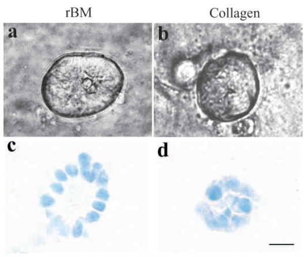
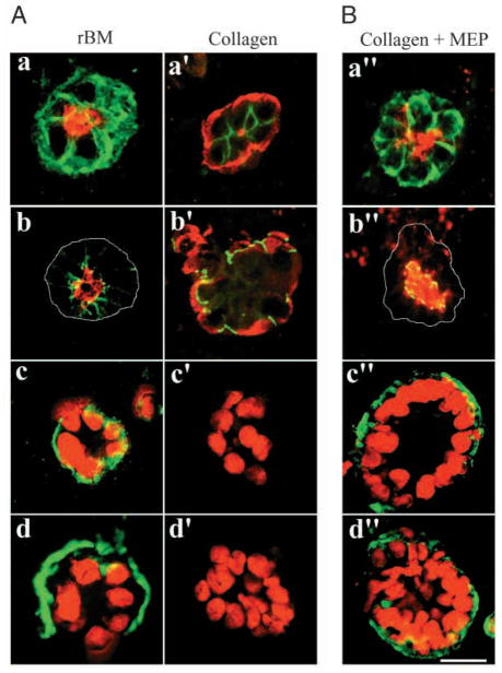
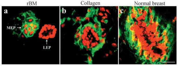
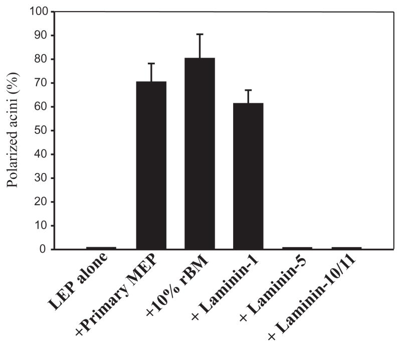
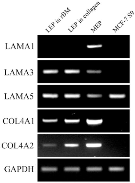
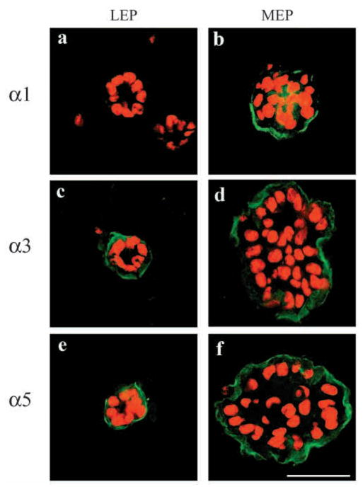
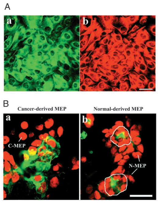
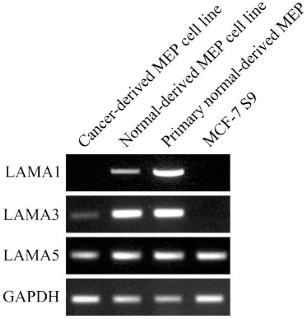
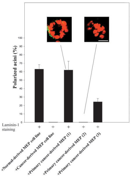
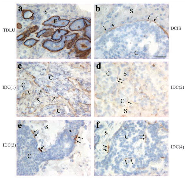
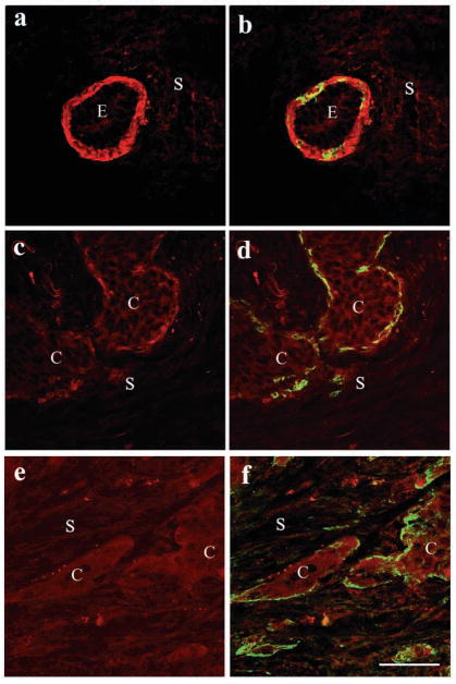
The signals that determine the correct polarity of breast epithelial structures in vivo are not understood. We have shown previously that luminal epithelial cells can be polarized when cultured within a reconstituted basement membrane gel. We reasoned that such cues in vivo may be given by myoepithelial cells. Accordingly, we used an assay where luminal epithelial cells are incorrectly polarized to test this hypothesis. We show that culturing human primary luminal epithelial cells within collagen-I gels leads to formation of structures with no lumina and with reverse polarity as judged by dual stainings for sialomucin, epithelial specific antigen or occludin. No basement membrane is deposited, and beta4-integrin staining is negative. Addition of purified human myoepithelial cells isolated from normal glands corrects the inverse polarity, and leads to formation of double-layered acini with central lumina. Among the laminins present in the human breast basement membrane (laminin-1, -5 and -10/11), laminin-1 was unique in its ability to substitute for myoepithelial cells in polarity reversal. Myoepithelial cells were purified also from four different breast cancer sources including a biphasic cell line. Three out of four samples either totally lacked the ability to interact with luminal epithelial cells, or conveyed only correction of polarity in a fraction of acini. This behavior was directly related to the ability of the tumor myoepithelial cells to produce alpha-1 chain of laminin. In vivo, breast carcinomas were either negative for laminin-1 (7/12 biopsies) or showed a focal, fragmented deposition of a less intensely stained basement membrane (5/12 biopsies). Dual staining with myoepithelial markers revealed that tumor-associated myoepithelial cells were either negative or weakly positive for expression of laminin-1, establishing a strong correlation between loss of laminin-1 and breast cancer. We conclude that the double-layered breast acinus may be recapitulated in culture and that one reason for the ability of myoepithelial cells to induce polarity is because they are the only source of laminin-1 in the breast in vivo. A further conclusion is that a majority of tumor-derived/-associated myoepithelial cells are deficient in their ability to impart polarity because they have lost their ability to synthesize sufficient or functional laminin-1. These results have important implications for the role of myoepithelial cells in maintenance of polarity in normal breast and how they may function as structural tumor suppressors.


Figures

Fig. 1
Fig. 1
Luminal epithelial cells form aberrant acini in collagen gels. Cells were embedded in either rBM (a,c) or hydrated collagen-I gels (b,d). (a,b) Phase-contrast microscopy revealed almost identical spherical structures in collagen-I and rBM albeit with no visible central lumen in spheres in collagen-I. (c,d) Haematoxylin staining of cryostat sections of the gels showed lumen formation only in rBM (bar, 25 μm).
Fig. 2
Fig. 2
(A) Luminal cells make inside-out acini in collagen. Luminal cells were double-stained either for (a) sialomucin (red) and ESA (green); (b) sialomucin (red) and occludin (green); (c) nuclear stain (red) and β4-integrin (green); or (d) nuclear stain (red) and type IV collagen (green). Spheres in collagen-I gel (a′,b′) exhibit reversed polarity compared with cells in rBM (a,b), do not target β4-integrin basolaterally (compare c and c′) and fail to deposit a basement membrane (compare d and d′). (B) Reversal of inside-out acini by addition of myoepithelial cells. In the presence of myoepithelial cells (a″-d″), the polarity is corrected as is the endogenous BM deposition and integrin targeting. (bar, 25 μm).
Fig. 3
Fig. 3
Luminal epithelial cells and myoepithelial cells sort themselves out inside rBM gels but form bilayered acini inside collagen gels. The spatial organization of myoepithelial cells in relation to luminal epithelial acinar backbones was analyzed in rBM (a) and hydrated collagen-I gel (b) and compared to that in normal breast (c). Gels were doublestained for Thy-1 to demonstrate myoepithelial cells (green) and propidium iodide to demonstrate nuclei (red). Note that whereas myoepithelial cells segregate from luminal epithelial acini in rBM, in vivo-like double-layered structures are formed in the collagen-I based acinus assay (bar, 25 μm).
Fig. 4
Fig. 4
Only laminin-1 and not laminin-5 and -10/11 can correctly polarize acini in collagen gels. Luminal epithelial cells without myoepithelial (LEP alone) had no correctly polarized acini. Addition of 10% rBM or pure laminin-1 reversed polarity to the same degree as normal-derived myoepithelial cells (MEP). Neither affinity purified laminin-5 nor laminin-10/11 possessed the ability to revert acinus polarity.
Fig. 5
Fig. 5
LAMA-1 chain is expressed only in myoepithelial cells. RT-PCR analysis of laminin α1 (LAMA1), laminin α3 (LAMA3), laminin α5 (LAMA5), type IV collagen α1 (COL4A1) and type IV collagen α2 (COL4A2) in luminal epithelial cells in rBM and collagen-I gel and in myoepithelial cells and MCF-7 cells. GAPDH is used as an internal control. Note that the only laminin chain that is not expressed in the pure luminal epithelial cultures is the α1-chain.
Fig. 6
Fig. 6
Luminal epithelial cells and myoepithelial cells can deposit both α3-chain and α5-chain of laminin but only myoepithelial cells can deposit laminin-α1 chain. Luminal epithelial cells (a,c,e) and myoepithelial cells (b,d,f) embedded in rBM were stained for laminin α1 (a,b; green), laminin α3 (c,d; green) and laminin α5 (e,f; green). Both luminal epithelial cells and myoepithelial cells deposit a basement membrane except for the lack of α1-chain deposition by the former. Nuclear stain, red. (bar, 25 μm).
Fig. 7
Fig. 7
Cancer-derived myoepithelial cells share many characteristics of normal myoepithelial cells but fail to reorient inside-out acini. (A) Isolation of cancer-derived myoepithelial cells expressing typical myoepithelial markers. Purified cancer-derived myoepithelial cells (a,b) stained with α-smooth muscle actin (green) and keratins (red) to document the concurrent myo- and epithelial phenotypes (bar, 50 μm). (B) Lack of reversal of inside-out acini by cancer-derived myoepithelial cells. Sections of (a) the acinus assay embedded with the cancer-derived myoepithelial cell line and (b) an immortalized, normal-derived myoepithelial cell line. The sections are double stained for the apical marker sialomucin (green) and the nuclear stain propidium iodide (red). Note the lack of polarization with the cancer myoepithelial cells (C-MEP) and the correctly polarized lumina (encircled) with the immortalized myoepithelial cells (N-MEP) (bar, 25μm).
Fig. 8
Fig. 8
The cancer-derived myoepithelial cell line lacks the expression of laminin-α1 chain only. RT-PCR on RNA extracted from cancer-derived myoepithelial cell line, immortalized, normal-derived myoepithelial cells (MEP), primary cultured MEP and MCF-7 S9 cells. The panel shows α1-chain (LAMA1), α3-chain (LAMA3), α5-chain (LAMA5) and GAPDH as an internal control. While the α3 mRNA was low in the cancer-derived MEP cell line, the αl-chain was the only differentially expressed chain between normal-derived and cancer-derived myoepithelial cells.
Fig. 9
Fig. 9
Only one of four cancer-derived myoepithelial cells can completely reverse acinus polarity in collagen gels. Frequency of correctly polarized acini in sections of collagen-I gels. Addition of cancer-derived myoepithelial cells from four different sources failed completely to revert acini in two instances, had a partial impact on reversion in one instance, and could revert in the fourth case. Inserts show staining for β4-integrin (green) counterstained with propidium iodide (red). Data represent mean (±s.e.m.) except for the primary cancer-derived MEP, which are means of triplicate determinations (±s.d.). (bar, 20 μm).
Fig. 10
Fig. 10
Reduced staining of laminin-1 in human breast cancer. Cryostat sections of terminal duct lobular unit (TDLU) (a), a ductal carcinoma in situ (DCIS) (b) and four different infiltrating ductal breast carcinomas (IDC) (c–f) stained with immunoperoxidase for laminin-1 and counterstained with hematoxylin. In the TDLU a strongly stained, continuous basement membrane is delineated at the stromal (S)-epithelial (E) interface (a). In carcinomas, laminin-1 staining is localized to the stromal (S)-cancer epithelial (C) interface, but is discontinuous and less pronounced. (bar, 25 μm).
Fig. 11
Fig. 11
Absence of, and reduced staining of laminin-1 in tumor-associated myoepithelial cells. Double-labeling immunofluorescence of a normal acinus in a TDLU (a,b) and two different carcinomas (c,d and e,f) to demonstrate laminin-1 in red (a,c,e) and in a coexposure with myoepithelial cells in green (b,d,f) as stained with keratin 17 (b,d) or maspin (f). The bottom panels illustrate the presence of myoepithelial cells at the stromal (S)-cancer epithelial (C) interface in total absence of laminin-1 staining. The middle panels show infiltrating ductal carcinoma with tumor-associated myoepithelial cells expressing a variable but generally weak staining for laminin-1 compared with a normal acinus in the top row. (bar, 50 μm).

Similar articles

Cited by

References

    1. Albrechtsen R, Nielsen M, Wewer U, Engvall E, Rouslahti E. Basement membrane changes in breast cancer detected by immunohistochemical staining for laminin. Cancer Res. 1981;41:5076–5081. - PubMed
    1. Band V, Zajchowski D, Kulesa V, Sager R. Human papilloma virus DNAs immortalize normal human mammary epithelial cells and reduce their growth factor requirements. Proc Natl Acad Sci USA. 1990;87:463–467. - PMC - PubMed
    1. Bani D, Riva A, Bigazzi M, Sacchi BT. Differentiation of breast cancer cells in vitro is promoted by the concurrent influence of myoepithelial cells and relaxin. Br J Cancer. 1994;70:900–904. - PMC - PubMed
    1. Briand P, Lykkesfeldt A. Long-term cultivation of a human breast cancer cell line, MCF-7, in a chemically defined medium. Effect of estradiol. Anticancer Res. 1986;6:85–90. - PubMed
    1. Clarke C, Titley J, Davies S, O’Hare MJ. An immunomagnetic separation method using superparamagnetic (MACS) beads for large-scale purification of human mammary luminal and myoepithelial cells. Epithelial Cell Biol. 1994;3:38–46. - PubMed

Publication types

MeSH terms