. 2002 Feb;111(1-2):99-113.
doi: 10.1016/s0925-4773(01)00614-1.

Evidence for coordinated interaction of cyclin D3 with p21 and cdk6 in directing the development of uterine stromal cell decidualization and polyploidy during implantation

Affiliations

Evidence for coordinated interaction of cyclin D3 with p21 and cdk6 in directing the development of uterine stromal cell decidualization and polyploidy during implantation

Jian Tan et al. Mech Dev. 2002 Feb.

Abstract

Uterine decidualization, characterized by stromal cell proliferation, and differentiation into specialized type of cells (decidual cells) with polyploidy, during implantation is critical to the pregnancy establishment in mice. The mechanisms by which the cell cycle events govern these processes are poorly understood. The cell cycle is tightly regulated at two particular checkpoints, G1-S and G2-M phases. Normal operation of these phases involves a complex interplay of cyclins, cyclin-dependent kinases (cdks) and cdk inhibitors (CKIs). We previously observed that upregulation of uterine cyclin D3 at the implantation site is tightly associated with decidualization in mice. To better understand the role of cyclin D3 in this process, we examined cell-specific expression and associated interactions of several cell cycle regulators (cyclins, cdks and CKIs) specific to different phases of the cell cycle during decidualization in mice. Among the various cell cycle molecules examined, coordinate expression and functional association of cyclin D3 with cdk4 suggest a role for proliferation and, that of cyclin D3 with p21 and cdk6 is consistent with the development of polyploidy during stromal cell decidualization.


Figures

Fig. 1
Fig. 1
In situ hybridization of cyclins D1-D3, cdk4 and cdk6 mRNAs in the mouse implantation sites. Frozen sections (10 μm) were mounted onto poly-L-lysine coated slides and fixed in 4% paraformaldehyde in PBS. The sections were hybridized with 35S-labeled sense or antisense riboprobes for 4 h at 45°C. RNase A resistant hybrids were detected after 5–7 days of autoradiography using Kodak NTB-2 liquid emulsion. Sections were post-stained lightly with hematoxylin and eosin. Dark-field photomicrographs of representative cross-sections are shown on day 5 at 09:00 h (D5am) and at 18:00 h (D5pm), and on days 6 (D6am) and 8 (D8am) at 09:00 h of pregnancy. Magnifications are shown at 40 × for days 5–6 pregnancy and at 20 × for day 8 of pregnancy. M, mesometrial pole; AM, antimesometrial pole; em, embryo; PDZ, primary decidual zone; SDZ, secondary decidual zone. Sections hybridized with the corresponding sense probes did not show any positive signals (data not shown).
Fig. 2
Fig. 2
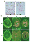
Analyses of cell cycle inhibitors at the implantation sites. (A) Immunohistochemical staining of p107 and p130 on D6 of pregnancy. Implantation sites at 09:00 h were fixed in formalin and paraffin embedded sections (6 μm) were mounted onto poly-L-lysine-coated slides. After deparaffinization and hydration, sections were incubated with primary antibody either for p107 or p130 in PBS for 17 h at 4°C. The photomicrographs of representative uterine sections are shown at 100 ×. Red nuclear staining indicates the localization of immunoreactive proteins. No immunostaining was noted when similar sections were incubated with the primary antibody pre-neutralized with excess antigen (data not shown). AM, antimesometrial pole; em, embryo; PDZ, primary decidual zone; SDZ, secondary decidual zone. (B) In situ hybridization of p21 and p27 mRNAs on D5, D6 and D8 of pregnancy. While dark-field photomicrographs of representative cross-sections on D5 and D6 are shown at 40 ×, representative cross-sections on D8 are shown at 20 ×. Sections hybridized with the corresponding sense probes did not show any specific signals (data not shown). M, mesometrial pole; AM, antimesometrial pole; em, embryo; PDZ, primary decidual zone; SDZ, secondary decidual zone.
Fig. 3
Fig. 3
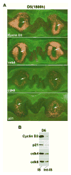
(A) In situ hybridization for cyclin D3, cdk4, cdk6 and p21 mRNAs in the implantation and inter-implantation sites on day 5 (18:00 h) of pregnancy. Dark-field photomicrographs of longitudinal uterine sections containing two implanting embryos are shown at 20 ×. Arrows indicate the location of embryo in the implantation site. (B) Western blot analysis of cyclin D3, p21, cdk4 and cdk6 proteins in the mouse uterus on D6 of pregnancy. Implantation (IS) and interimplantation (Int-IS) sites were collected separately, and extracted and fractionated by 10% SDS-PAGE. Immunoblotting was performed using primary antibodies specific to cyclin D3, p21, cdk4 or cdk6. The primary antibodies and the protocol used for immunoblotting are described in Materials and Methods. For control experiments, primary antibodies pre-neutralized with 250-fold molar excess of corresponding antigenic peptide were used (data not shown).
Fig. 4
Fig. 4
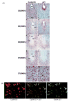
(A) Immunohistochemical staining of cyclin D3 and p21 in the mouse uterus during decidualization. Mouse uteri collected during pregnancy on D5 at 09:00 and 18:00 h, and D6 and D7 at 09:00 h were analyzed. Formalin-fixed paraffin embedded adjacent sections (6 μm) collected from an implantation site were mounted onto poly-L-lysine-coated slides. After deparaffinization and hydration, sections were incubated with primary antibody either for cyclin D3 or p21 in PBS for 17 h at 4°C. The photomicrographs of representative uterine sections are shown with bars, 100 μm. Red nuclear staining indicates the localization of immunoreactive proteins. Mono- and bi-nucleated cells are indicated by arrowheads and arrows, respectively. No immunostaining was noted when similar sections were incubated with the primary antibody pre-neutralized with excess antigen (data not shown). M, mesometrial pole; AM, antimesometrial pole; em, embryo; le, luminal epithelium; ds, decidualizing stroma,; PDZ, primary decidual zone; SDZ, secondary decidual zone. (B) Dual immunofluorescence study for cyclin D3 and p21 on day 6 of pregnancy at the antimesometrial pole of the implantation site. Uterine sections were stained with Cy3 (red) or FITC (green) tagged secondary antibody for cyclin D3 and p21, respectively. A superimposed picture at the middle, showing cells highlighted with yellow color, actually represents the expression of both cyclin D3 and p21. Arrows or arrowhead represent mono- or bi-nucleated polyploid cells, respectively.
Fig. 5
Fig. 5
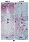
Immunohistochemical staining of cdk4 and cdk6 in the mouse uterus during decidualization on D6 of pregnancy. The photomicrographs of representative uterine sections are shown for cdk4 (a,c) and cdk6 (b,d) in locations of mesometrial (a,b) and antimesometrial (c,d) regions of the decidual bed at 100 ×. Red staining indicates the localization of immunoreactive proteins. No immunostaining was noted when similar sections were incubated with the primary antibody pre-neutralized with excess antigen (data not shown). M, mesometrial pole; AM, antimesometrial pole; em, embryo; PDZ, primary decidual zone; SDZ, secondary decidual zone.
Fig. 6
Fig. 6
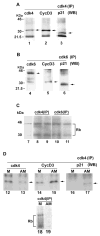
Analyses of the associated complex and the functional activity formed by cdk4 or cdk6 together with cyclin D3 and p21. (A) Complex with cdk4: uterine implantation sites on D6 of pregnancy were extracted and subjected to immunoprecipitation using cdk4 polyclonal antibody. Immunoprecipitates were separated on 10% SDS-PAGE gel, transferred to nitrocellulose membrane and then detected by Western blotting using antibodies for cdk4, cyclin D3 or p21. The bands specific to cdk4, cyclin D3 and p21 are indicated by arrows in lanes 1, 2 and 3, respectively. (B) Complex with cdk6: uterine implantation sites on D6 of pregnancy were extracted, immunoprecipitated by cdk6 polyclonal antibody, separated on 10% SDS-PAGE and analyzed by Western blotting using primary antibodies for cdk6, cyclin D3 and p21. The bands specific to cdk6, cyclin D3 and p21 are indicated by arrows in lanes 4, 5 and 6, respectively. (C) Kinase activities in cdk4- or cdk6-associated complex at the site of implantation on day 6 of pregnancy: Immunoprecipitates as obtained from two independent samples of tissue extracts of D6 implantation sites using cdk4 (lanes 8 and 9) or cdk6 (lanes 10 and 11) antibodies were subjected to kinase assay using [γ-32P]ATP and a protein substrate (mouse recombinant Rb fusion protein) as described in Materials and Methods. The phosphorylated proteins were separated by 10% SDS-PAGE, dried and subjected to autoradiography. The recombinant Rb was run on the same gel in parallel and visualized by Coomasie blue staining (lane 7). Two phosphorylated protein bands corresponding to Rb were detected as 45 and 30 kDa. (D) Analyses of cdk4-associated immunocomplex and its kinase activity in the separated antimesometrial (AM) and mesometrial (M) deciduomal tissues obtained on day 7 after induction of artificial decidualization on day 4 of pseudopregnancy. The bands specific to cdk4, cyclin D3 and p21 are indicated by arrows (lanes 12–17). Lanes 18 and 19 are shown for cdk4-associated kinase activity in the mesometrial (M) and antimesometrial (AM) deciduomal tissues, respectively.
Fig. 7
Fig. 7
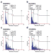
DNA content and the cell cycle distribution of uterine antimesometial stromal cells at the implantation sites. Formalin-fixed paraffin embedded sections (6 μm) of implantation sites on D5–D7 of pregnancy at indicated times were subjected to Feulgen staining and analyzed for measurements of DNA content by CAS-200 imaging system as described in Materials and Methods. In order to demarcate the location of the antimesometial decidual bed, adjacent sections were examined in parallel by immuonohistochemical staining for cyclin D3 and p21 as shown in Fig. 3A. On each day of pregnancy, four different implantation sites on duplicate adjacent sections were analyzed. In average, 500–600 nuclei were analyzed for each implantation site. The DNA content for diploid nuclei was optimized with non-differentiated stromal cells on day 4 of pregnancy. The percent of cell cycle distribution was calculated as mean ± SEM. The colored bars represent several multiples of normal haploid DNA content (C).
Fig. 8
Fig. 8
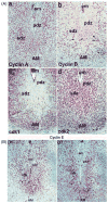
(A) Expression of cyclin A, cyclin B, cdk1 and cdk2 in the uterus on D6 of pregnancy. (B Expression of cyclin E on days 6 and 7 of pregnancy. Immunohistochemical staining on formalin-fixed paraffin sections (6 μm) of implantation sites is shown at 100 ×. The photomicrographs of representative uterine sections are shown mostly for the antimesometrial region of the decidual bed. Red staining indicates the localization of immunoreactive proteins. No immunostaining was noted when similar sections were incubated with the primary antibody pre-neutralized with excess antigen (data not shown). Arrows indicate polyploid cells. M, mesometrial pole; AM, antimesometrial pole; em, embryo; PDZ, primary decidual zone; SDZ, secondary decidual zone.
Fig. 9
Fig. 9
A potential model for stromal cell transition from the mitotic to the endocycle pathway for polyploidization. This model describes presumed functional association of the G1 phase cell regulators (cyclin D3, p21, cdk4, cdk6, cyclin E and cdk2) and that of the S-G2-M phases (cyclin A, cyclin B, cdk1 and cdk2) with respect to cell cycle events associated with the switch from mitotic cycle to endocycle during stromal cell decidualization. The absence of cyclin B and cdk1 is speculated to participate with the onset of first endocycle.

Similar articles

Cited by

References

    1. Ansell JD, Barlow PW, McLaren A. Binucleate and polyploid cells in the decidua of the mouse. J Embryol Experi Morphol. 1974;31:223–227. - PubMed
    1. Bamberger AM, Sudahl S, Bamberger CM, Schulte HM, Lo ning T. Expression patterns of the cell-cycle inhibitor p27 and the cell cycle promoter cyclin E in the human placenta throughout gestation: Implications for the control of proliferation. Placenta. 1999;20:401–406. - PubMed
    1. Das SK, Lim H, Paria BC, Dey SK. Cyclin D3 in the mouse uterus is associated with the decidualization process during early pregnancy. J Mol Endocrinol. 1999;22:91–101. - PubMed
    1. Deng C, Zhang P, Harper JW, Elledge SJ, Lader P. Mice lacking p21Cip1/Waf1 undergo normal development, but are defective in G1 checkpoint control. Cell. 1995;82:675–684. - PubMed
    1. Dey SK. Implantation. In: Adashi EY, Rock JA, Rosenwaks Z, editors. Reproductive Endocrinology, Surgery and Technology. Lippincott-Raven Publishers; New York: 1996. pp. 421–434.

Publication types

MeSH terms

LinkOut - more resources