. 2002 Jan;109(2):183-92.
doi: 10.1172/JCI11703.

Beta2 integrins are required for skin homing of primed T cells but not for priming naive T cells

Affiliations

Beta2 integrins are required for skin homing of primed T cells but not for priming naive T cells

Stephan Grabbe et al. J Clin Invest. 2002 Jan.

Abstract

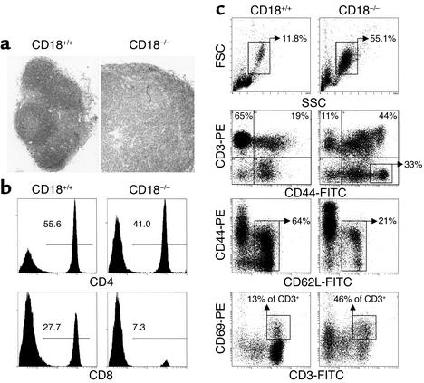
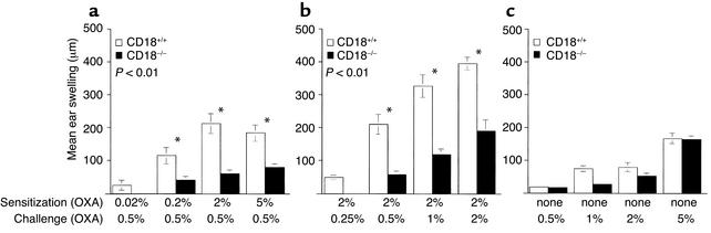
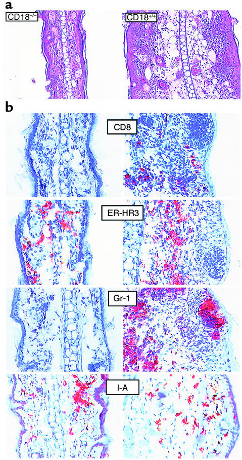
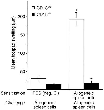
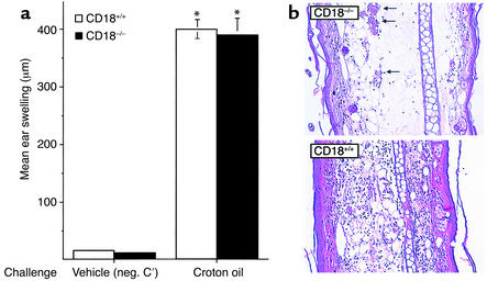
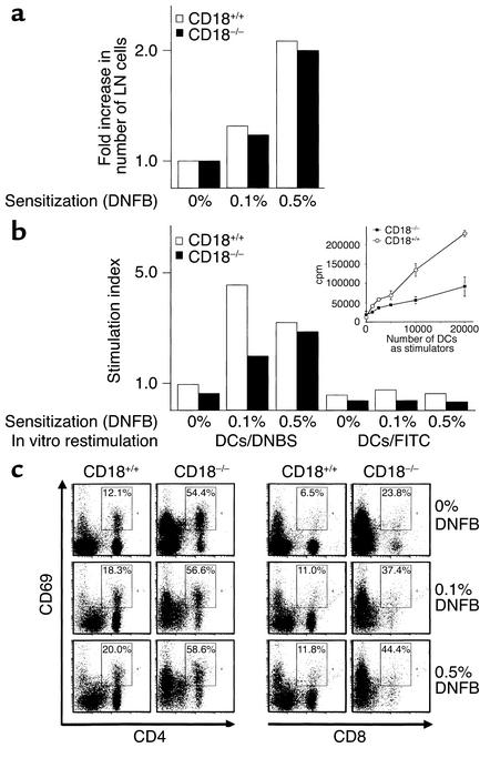
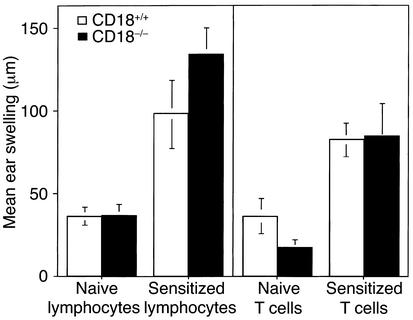
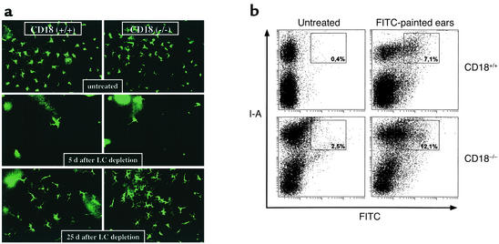
Beta2 integrins are of critical importance for leukocyte extravasation through vascular endothelia and for T cell activation. To elucidate the role of beta2 integrins in T cell-mediated immune responses, allergic contact dermatitis (ACD), irritant dermatitis, and delayed-type hypersensitivity (DTH) were assessed in mice lacking the beta2 integrin subunit, CD18. ACD and DTH responses, but not edema formation, were severely suppressed in CD18(-/-) mice. Extravasation of CD18(-/-) T cells into eczematous skin lesions was greatly impaired, whereas migration of Langerhans cell precursors and dendritic cells was normal in CD18(-/-) mice. CD18(-/-)lymph nodes (LNs) contained an abnormal population of CD3(-)CD44(high) lymphocytes and showed evidence of widespread T cell activation. T cells from regional LNs of sensitized CD18(-/-) mice proliferated in response to hapten challenge, and subcutaneous injection of sensitized syngeneic LN cells directly into ears of hapten-challenged naive recipients restored the defective ACD in CD18(-/-) mice, suggesting that CD18 is not required for priming of naive T cells but is indispensable for T cell extravasation. Thus, a dysfunction of T cells, in addition to granulocytes, may contribute to the pathophysiology of leukocyte adhesion deficiency type I, which arises from mutations in the human CD18 gene.


Figures

Figure 1
Figure 1
Phenotype of peripheral LNs in CD18–/– mice. (a) Histopathology of LNs. Whereas lymph follicles are present in CD18+/+ mice, the architecture of the LN is dissolved in CD18–/– mice, and the LNs are greatly enlarged. Original magnification, ×100. (b) One-color flow cytometry of LN cells. (c) Two-color flow cytometry of LN cells. Numbers indicate percentage of cells in the respective box or quadrant. FSC, forward scatter; SSC,sideward scatter.
Figure 2
Figure 2
ACD and irritant dermatitis to epicutaneous application of OXA in CD18–/– mice. Mice were sensitized to OXA by epicutaneous application of various concentrations of OXA as described in Methods. Six days later, mice were OXA-challenged at one ear to induce ACD, and ear swelling was determined 30 hours later. (a) ACD, titration of sensitization dose. (b) ACD, titration of challenge dose. (c) Irritant dermatitis to epicutaneous application of OXA.
Figure 3
Figure 3
ACD in CD18–/– mice. (a) Histology (hematoxylin and eosin staining; original magnification, ×200) and (b) immunohistochemistry (original magnification, ×400) of OXA-challenged ears from sensitized CD18–/– and WT control mice, obtained 30 hours after challenge with 0.5% OXA. Antibodies were used to detect the following leukocyte subsets: cytotoxic T cells (detected with anti-CD8), monocytes and macrophages (detected with anti–ER-HR3), granulocytes (detected with anti–Gr-1), and activated macrophages and DCs (detected with anti–I-A).
Figure 4
Figure 4
DTH in CD18–/– mice. Alloantigen-specific DTH was elicited by subcutaneous injection of 108 spleen cells from BALB/c (H-2d) mice into footpads of allosensitized CD18–/– or control (H-2b) mice, and footpad swelling responses were determined 24 hours later. *P < 0.001. Neg. C′, negative control.
Figure 5
Figure 5
Irritant dermatitis in CD18–/– mice. Irritant dermatitis was elicited by application of 10 μl 0.8% croton oil to both sides of one ear, and measurement of ear swelling responses (a) and histological examination (b) were performed 18 hours later. Arrows indicate dilated blood vessels with endothelium-associated leukocytes, some of which appear to have extravasated. Original magnification, ×200. *P = not significant.
Figure 6
Figure 6
Hapten-specific activation of regional LN cells. Regional LNs were obtained from DNFB-sensitized mice 3 days after sensitization. (a) Fold increase in LN cells after sensitization with either vehicle only (0% DNFB), 0.1%, or 0.5% DNFB. (b) Hapten-specific proliferation of LN cells in response to either the specific hapten, DNBS, or a control hapten, FITC. Thy-1.2+ T cells were generated from LN cells and cultured together with hapten-coupled DCs for 72 hours, and then pulsed with 3H for an additional 18 hours. Due to the different proliferative capacities of CD18–/– and CD18+/+ T cells, a proliferation index was calculated as cpm after stimulation with hapten-exposed DCs, divided by cpm after stimulation with untreated DCs. A proliferation index greater than 1 indicates hapten-specific T cell proliferation. Inset: Allogeneic mixed lymphocyte reaction with purified T cells from CD18–/– or CD18+/+ mice as responders, and various numbers of allogeneic DCs (156–20,000) as stimulators. (c) Hapten-specific T cell activation as measured by upregulation of the activation marker CD69. Numbers in boxes indicate the percentage of CD69-expressing cells among the CD4+ and CD8+ T cells.
Figure 7
Figure 7
ACD in CD18–/– mice after subcutaneous injection of syngeneic LN cells. Left panel: CD18–/– and WT mice were OXA-sensitized, and regional LNs were obtained. LN cells (2.5 × 10⁶) from sensitized or naive CD18–/– mice were injected into ears of autologous OXA-challenged CD18–/– mice, and 2.5 × 10⁶ LN cells from sensitized or naive WT mice were injected into ears of autologous OXA-challenged WT mice. Ear swelling responses were determined after 32 hours. P < 0.05 compared with transfer of naive LN cells of the same mouse strain. Right panel: The experiment was performed in a similar fashion as in the left panel, except that purified T cells from CD18–/– mice were transferred in all cases. P < 0.05 compared with transfer of naive LN cells of the same mouse strain. No significant differences in ear swelling were present between ears injected with whole LN cells or purified T cells.
Figure 8
Figure 8
Accumulation of 51Cr-labeled T cells and DCs at skin sites of ACD. Mice were sensitized and challenged with the hapten OXA. T cells and DCs were generated as described in Methods, labeled with 51Cr, and injected intravenously 24 hours after hapten challenge. Six hours later, ears and internal organs were harvested and 51Cr was counted. Data represent percentage of incorporated 51Cr in the ear in relation to the combined radioactivity uptake in lungs, liver, and spleen. *P < 0.01 for WT versus CD18–/– T cells; P = not significant for all other groups.
Figure 9
Figure 9
LCs in WT and CD18–/– mice. (a) Mice that were either left untreated, or were treated with topical mometasone-17-(2-furoate) 5 days or 25 days prior to assessment of LC density. Ear skin was obtained, and epidermal sheets were prepared and stained for I-A+ epidermal cells, as described in Methods. (b) Ears were treated with the fluorescent hapten FITC. Regional LNs were prepared 18 hours later, and LN cells were stained with phycoerythrin-labeled anti–I-A antibodies and subjected to flow cytometry. LCs were gated based on FSC/SSC characteristics. Numbers in boxes indicate the percentage of double positive cells.

Similar articles

Cited by

References

    1. Hynes RO. Integrins: versatility, modulation, and signaling in cell adhesion. Cell. 1992;69:11–25. - PubMed
    1. Issekutz TB. Lymphocyte homing to sites of inflammation. Curr Opin Immunol. 1992;4:287–293. - PubMed
    1. Luscinskas FW, Lawler J. Integrins as dynamic regulators of vascular function. FASEB J. 1994;8:929–938. - PubMed
    1. Benimetskaya L, et al. Mac-1 (CD11b/CD18) is an oligodeoxynucleotide-binding protein. Nat Med. 1997;4:414–420. - PubMed
    1. El Ghmati SM, van Hoeyveld EM, van Strijp JAG, Ceuppens JL, Stevens EAM. Identification of haptoglobin as an alternative ligand for CD11b/CD18. J Immunol. 1996;156:2542–2552. - PubMed

Publication types