. 2002 Jan;109(2):251-9.
doi: 10.1172/JCI14040.

A chemokine receptor CCR-1 antagonist reduces renal fibrosis after unilateral ureter ligation

Affiliations

A chemokine receptor CCR-1 antagonist reduces renal fibrosis after unilateral ureter ligation

Hans-Joachim Anders et al. J Clin Invest. 2002 Jan.

Abstract

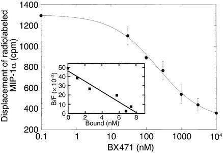
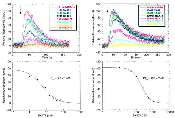
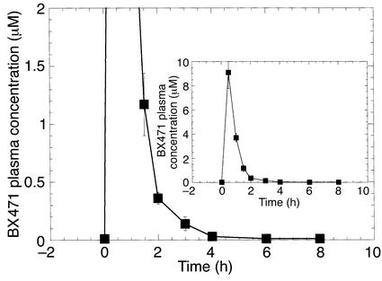
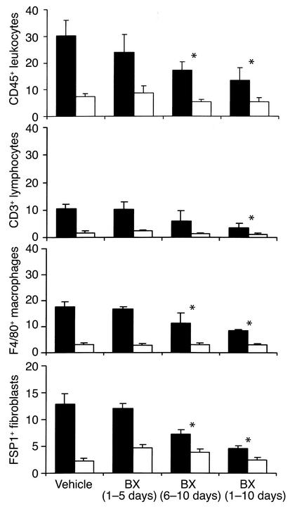
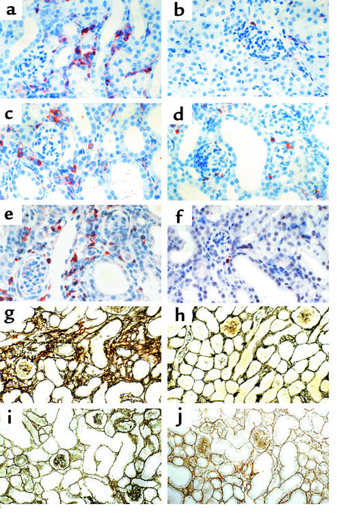
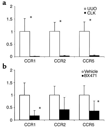
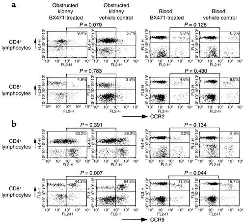
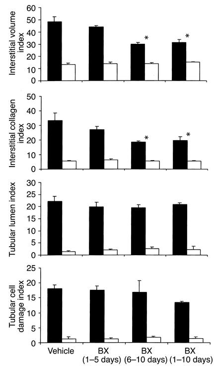
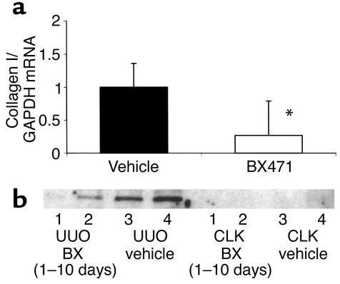
The expression of chemokines and their receptors is thought to contribute to leukocyte infiltration and progressive renal fibrosis after unilateral ureter obstruction (UUO). We hypothesized that blocking the chemokine receptor CCR1 using the nonpeptide antagonist BX471 could reduce leukocyte infiltration and renal fibrosis after UUO. UUO kidneys from BX471-treated mice (day 0-10 and day 6-10) revealed a 40-60% reduction of interstitial macrophage and lymphocyte infiltrate compared with controls. Treated mice also showed a marked reduction of CCR1 and CCR5 mRNA levels, and FACS analysis showed a comparable reduction of CD8+/CCR5+ T cells. Markers of renal fibrosis, such as interstitial fibroblasts, interstitial volume, mRNA and protein expression for collagen I, were all significantly reduced by BX471-treatment compared with vehicle controls. By contrast treatment was ineffective when the drug was supplied only from days 0 to 5. In summary, blockade of CCR1 substantially reduces cell accumulation and renal fibrosis after UUO. Most interestingly, late onset of treatment is also effective. We therefore conclude that CCR1 blockade may represent a new therapeutic strategy for reducing cellular infiltration and renal fibrosis as major factors in the progression to end-stage renal failure.


Figures

Figure 1
Figure 1
Binding of BX471 to murine CCR1. The CCR1 antagonist, BX471, displaces radiolabeled MIP-1α from murine CCR1. HEK cells transfected with murine CCR1 were incubated with 125I-MIP-1α/CCL3 in the presence of increasing concentrations of BX471. The binding reactions were terminated by centrifugation of cells as described previously (14). Binding shown represents specific binding from a typical experiment (n = 3). Nonspecific binding was 10% of total 125I-MIP-1α/CCL3 added. Inset shows the Scatchard plot of the displacement data.
Figure 2
Figure 2
Cytosolic Ca2+ measurements in HEK 293 cells. BX471 inhibited the ability of MIP-1α/CCL3 to increase Ca2+ transients in HEK 293 cells expressing human and murine CCR1. Fluo-3–loaded cells were pretreated with increasing concentrations of BX471 for 15 minutes and then stimulated with the CCR1 agonist, MIP-1α/CCL3. The changes in fluorescence representing the changes in Ca2+ concentration were measured as indicated under Methods.
Figure 3
Figure 3
Pharmacokinetics of BX471 in mice. Plasma concentrations of BX471 following subcutaneous dosing in mice. Male mice (n = 4) received a single subcutaneus dose (20 mg/kg) of BX471 in 40% cyclodextrin. Blood plasma levels of BX471 were measured as described previously (9). Data are the mean ± SEM.
Figure 4
Figure 4
Accumulation of interstitial cells after UUO. Quantitative analysis of cell infiltration 10 days after UUO in obstructed (black bars) and contralateral unobstructed kidneys (white bars). Values are means ± SD of cell counts per hpf. Note that treatment with BX471 for 10 days as well as from days 6–10 reduced infiltration of CD45+ leukocytes, CD3+ lymphocytes, F4/80+ macrophages, and FSP1+ fibroblasts compared with vehicle controls. BX471 given from day 0–5 had no effect. * P < 0.05.
Figure 5
Figure 5
Leukocyte infiltration and renal fibrosis after UUO histology. Cortical renal sections were stained for CD45+ leukocytes. At 10 days after UUO (a) leukocytes accumulated in the peritubular interstitium in obstructed kidneys compared with unobstructed control kidneys (b). Treatment with BX471 from day 6 to 10 (c), as well as treatment for 10 days (d), reduced infiltration of CD45-positive leukocytes in UUO kidneys compared with a and b. No reduction was noted in UUO kidneys of mice treated with BX471 from days 0 to 5 (not shown). The accumulation of FSP1-positive fibroblasts in the peritubular interstitium 10 days after UUO (e) was reduced when BX471was given for 10 days (f) and from day 6 to 10 (latter not shown). Cortical renal sections were also silver stained to assess interstitial volume and fibrous tissue deposition. Note a marked widening of the interstitial space with deposition of fibrous tissue in areas of dilated tubules in obstructed kidneys at 10 days (g) compared with unobstructed control kidneys (h) of vehicle controls. Treatment with BX471 from days 6 to 10 reduced interstitial volume and matrix deposition (i), as well as BX471 treatment for 10 days (j), compared with g. No reduction was noted in UUO kidneys of mice treated with BX471 at days 0–5 (not shown). Tubular dilatation and tubular epithelial cell damage remained unchanged in all groups (original magnification ×400).
Figure 6
Figure 6
Renal chemokine receptor expression after UUO. Real-time RT-PCR was performed using total renal RNA of obstructed (UUO) and contralateral (CLK) kidneys of four to six mice 10 days after ureter ligation. Levels of mRNA expression for CCR1, CCR2, and CCR5 are expressed in relation to renal GAPDH mRNA expression as indicated in Methods. The expression in UUO kidneys of control mice is set as 1. (a) A marked increase of mRNA for all three receptors was noted in UUO kidneys compared with CLK of vehicle controls. (b) Treatment with BX471 for 10 days resulted in a significant decrease of CCR1 and CCR5 mRNA expression (P = 0.005 and P = 0.03, respectively) compared with UUO kidneys of vehicle controls. For CCR2 the difference was not significant (P = 0.07). *P < 0.05.
Figure 7
Figure 7
CCR2 and CCR5 expression on infiltrating leukocytes. Flow cytometric analysis of CCR2 (a) and CCR5 (b) expression on T cells isolated from obstructed kidneys and blood of BX471-treated mice and vehicle controls. Treatment with BX471 results in a significant decrease of CCR5-positive CD8+ lymphocytes infiltrating the obstructed kidneys and, to a lower extent, also in the peripheral blood. No significant changes were seen for CCR5-positive CD4 cells or CCR2-positive cells in kidneys or blood from BX471- or vehicle-treated mice. Dot blots are representative for four to six separate sets of experiments. Numbers given are means of five to nine mice analyzed in each group.
Figure 8
Figure 8
Histopathological damage after UUO: morphometry data. Morphometric analysis of cortical renal sections 10 days after UUO in obstructed kidneys (black bars) and contralateral unobstructed kidneys (white bars). Values are means ± SD of cell counts per hpf. Note that treatment with BX471 for 10 days as well as from days 6 to 10 significantly reduced the interstitial volume index and collagen index compared with vehicle controls. BX471 given from day 0 to 5 had no effect on interstitial volume and collagen index. The indices of tubular lumen and tubular cell damage were not affected by either treatment regimen.
Figure 9
Figure 9
Collagen I mRNA and protein expression. (a) Renal collagen mRNA expression was determined by real-time RT-PCR. Collagen Iα1 mRNA was analyzed as the ratio to GAPDH template and compared with the unligated kidney. Collagen I expression of the vehicle group is set as 1. A significant decrease of collagen Iα1 mRNA was found in BX471-treated animals (treatment from day 0 to 10) compared with vehicle-treated mice. (b) Levels of collagen I were determined by Western blot analysis. Protein extractions of two obstructed (UUO) and two unobstructed contralateral kidneys (CLK) of BX471-treated mice (lanes 1 and 2) were compared with vehicle controls (lanes 3 and 4). Note an increase of collagen I in UUO compared with CLK kidneys. Treatment with BX471 for 10 days reduced collagen I protein expression in UUO kidneys compared with control UUO kidneys.

References

    1. Becker GJ, Hewitson TD. The role of tubulointerstitial injury in chronic renal failure. Curr Opin Nephrol Hypertens. 2000;9:133–138. - PubMed
    1. Zeisberg M, Strutz F, Muller GA. Renal fibrosis: an update. Curr Opin Nephrol Hypertens. 2001;10:315–320. - PubMed
    1. Zlotnik A, Yoshie O. Chemokines: a new classification system and their role in immunity. Immunity. 2000;12:121–127. - PubMed
    1. Segerer S, Nelson PJ, Schlöndorff D. Chemokines, chemokine receptors, and renal disease: from basic science to pathophysiologic and therapeutic studies. J Am Soc Nephrol. 2000;11:152–176. - PubMed
    1. Segerer S, et al. Expression of chemokines and chemokine receptors during human renal transplant rejection. Am J Kidney Dis. 2001;37:518–531. - PubMed

Publication types

MeSH terms