. 2002 Jan 21;195(2):189-200.
doi: 10.1084/jem.20011571.

Vav3 modulates B cell receptor responses by regulating phosphoinositide 3-kinase activation

Affiliations

Vav3 modulates B cell receptor responses by regulating phosphoinositide 3-kinase activation

Kazunori Inabe et al. J Exp Med. .

Abstract

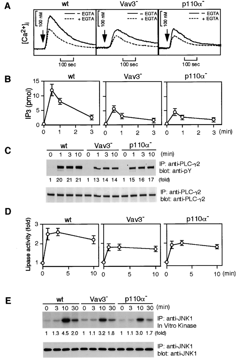
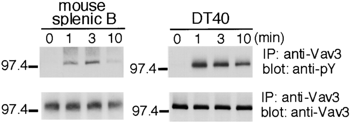
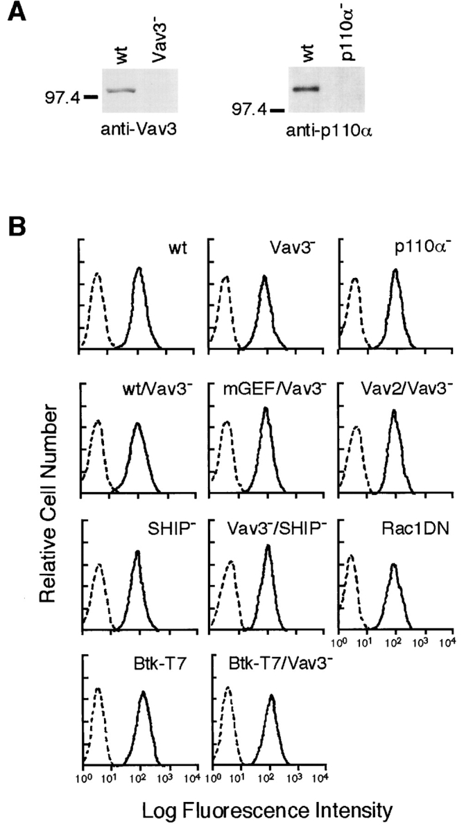
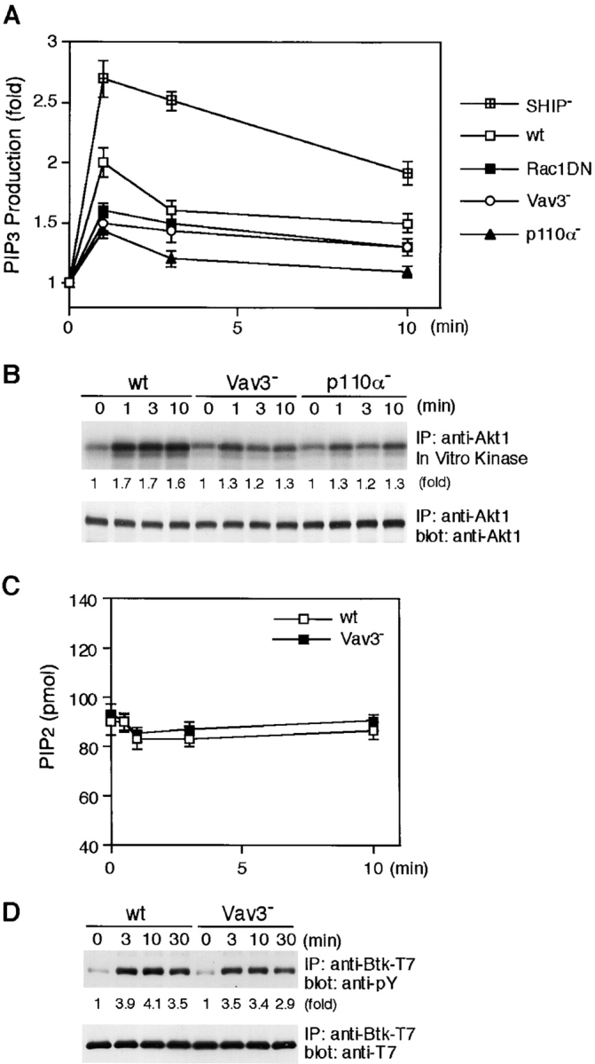
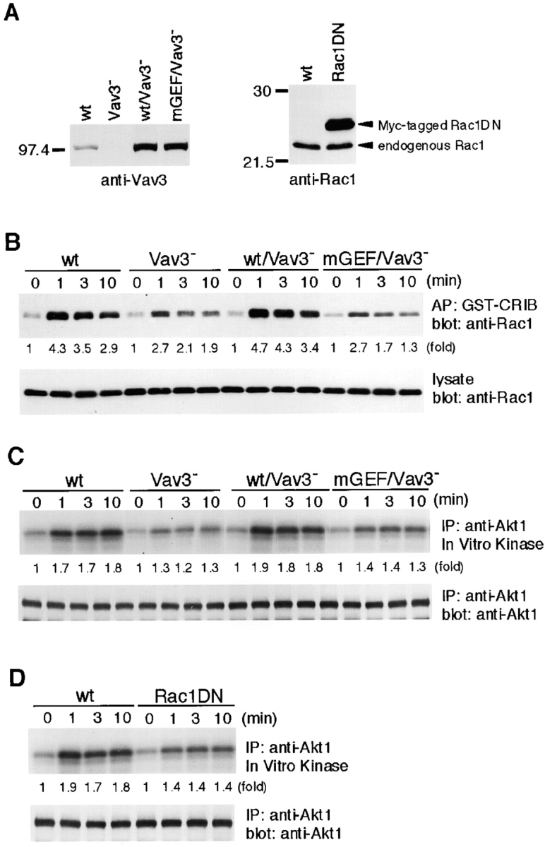
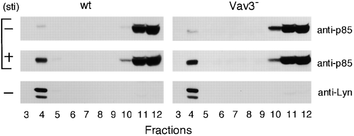
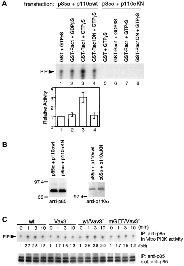
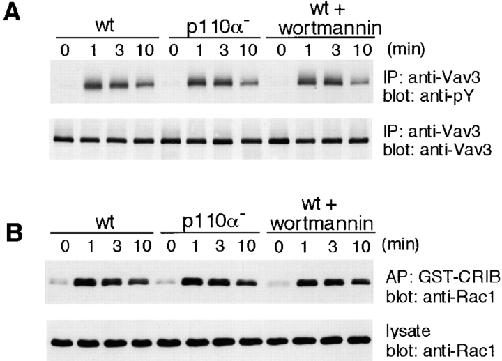
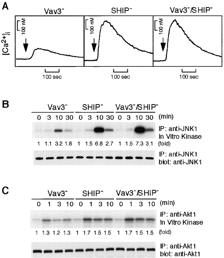
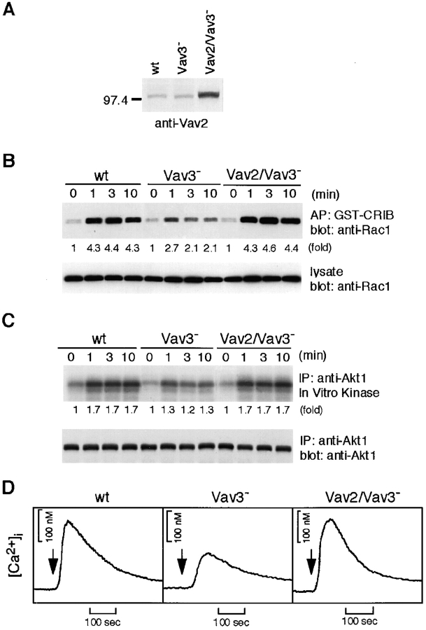
To elucidate the mechanism(s) by which Vav3, a new member of the Vav family proteins, participates in B cell antigen receptor (BCR) signaling, we have generated a B cell line deficient in Vav3. Here we report that Vav3 influences phosphoinositide 3-kinase (PI3K) function through Rac1 in that phosphatidylinositol-3,4,5-trisphosphate (PIP3) generation was attenuated by loss of Vav3 or by expression of a dominant negative form of Rac1. The functional interaction between PI3K and Rac1 was also demonstrated by increased PI3K activity in the presence of GTP-bound Rac1. In addition, we show that defects of calcium mobilization and c-Jun NH2-terminal kinase (JNK) activation in Vav3-deficient cells are relieved by deletion of a PIP3 hydrolyzing enzyme, SH2 domain-containing inositol polyphosphate 5'-phosphatase (SHIP). Hence, our results suggest a role for Vav3 in regulating the B cell responses by promoting the sustained production of PIP3 and thereby calcium flux.


Figures

Figure 3.
Figure 3.
Characterization of Vav3- and PI3K p110α-deficient DT40 B cells. Cells were stimulated with M4 (4 μg/ml) for indicated time periods and then subjected to the following analyses. (A) Calcium mobilization. Intracellular free calcium levels in Fura-2-loaded cells were monitored by a fluorescence spectrophotometer. Calcium release from intracellular calcium stores was measured in the presence of 1 mM EGTA (dotted line). wt, wild-type. (B) IP3 generation. Soluble IP3 was extracted from 2 × 10⁶ cells and subjected to a Biotrak competitive binding assay system. The results (2 × 10⁶ cell equivalents per point) were shown by mean ± standard error of three independent experiments. (C) Tyrosine phosphorylation of PLC-γ2. Immunoprecipitation (IP) was performed with anti-PLC-γ2 Abs. The blots (5 × 10⁶ cell equivalents per lane) were stained with 4G10 mAb (top). The blots were stripped and reprobed with anti-PLC-γ2 Abs (bottom). (D) BCR-induced PLC-γ2 activation. PLC-γ2 was immunoprecipitated from lysates (2 × 10⁷ cell equivalents per point), and in vitro phospholipase activity was measured as described in Materials and Methods. The results were shown by mean ± standard error of three independent experiments. (E) BCR-induced JNK1 activation. Lysates from 5 × 10⁶ cells were immunoprecipitated with anti-JNK1 mAb, and the resulting immunoprecipitates were divided. Half of them was used for Western blot analysis using anti-JNK1 mAb (bottom). The remaining half was used for in vitro kinase assay using GST-c-Jun as an exogenous substrate. The kinase reaction products were resolved on 12.5% SDS-PAGE, and their phosphorylation was quantified by autoradiography (top).
Figure 1.
Figure 1.
Vav3 is tyrosine phosphorylated after antigen-receptor ligation in B lymphocytes. Primary B cells purified from mouse spleen (107 cells per sample, left panels) and chicken DT40 B cells (5 × 10⁶ cells per sample, right panels) were incubated with 15 μg/ml polyclonal F(ab′)2 anti–mouse IgM and 4 μg/ml monoclonal anti–chicken IgM, M4, respectively. After stimulation, immunoprecipitates with anti-Vav3 Abs were separated on 7.5% SDS-PAGE and analyzed by Western blotting with 4G10 mAb (top) and anti-Vav3 Abs (bottom). IP, immunoprecipitation.
Figure 2.
Figure 2.
Generation of Vav3- and PI3K p110α-deficient DT40 B cells. (A) Cell lysates were separated on 7.5% SDS-PAGE and analyzed by Western blotting with anti-Vav3 Abs (left) or anti-p110α Abs (right). (B) Cell surface expression of BCR on wild-type and mutant DT40 cells used in this study: wt, parental DT40 cells; Vav3, Vav3-deficient cells; p110α, p110α-deficient cells; wt/Vav3, Vav3-deficient cells expressing wild-type Vav3; mGEF/Vav3, Vav3-deficient cells expressing GEF mutant Vav3; Vav2/Vav3, Vav3-deficient cells overexpressing Vav2; SHIP, SHIP-deficient cells; Vav3/SHIP, Vav3/SHIP double-deficient cells; Rac1DN, cells expressing dominant negative Rac1 (Rac1N17); Btk-T7, cells expressing Btk-T7; Btk-T7/Vav3, Vav3-deficient cells expressing Btk-T7. Unstained cells were used as negative controls (dotted line).
Figure 4.
Figure 4.
Impairment of BCR-induced PI3K signaling in the absence of Vav3. (A) PIP3 generation. Cells loaded with [32P]orthophosphate (107 cells per condition) were stimulated with M4 (4 μg/ml) for indicated time periods. PIP 3 was extracted and analyzed as described in Materials and Methods. Fold increases of PIP3 level normalized to total phospholipids after M4 stimulation were shown. These results were shown by mean ± standard error of three independent experiments. wt, wild-type. (B) BCR-induced Akt activation. Lysates from 5 × 10⁶ cells were immunoprecipitated with anti-Akt1 Abs, and the resulting immunoprecipitates were divided. Half of them was used for Western blot analysis using anti-Akt1 Abs (bottom). The remaining half was used for in vitro kinase assay using histone H2B as an exogenous substrate. The kinase reaction products were resolved on 15% SDS-PAGE, and their phosphorylation was quantified by autoradiography (top). IP, immunoprecipitation. (C) PIP2 levels. Wild-type and Vav3-deficient DT40 cells (2 × 10⁶) were stimulated with M4 (4 μg/ml) for indicated times and then processed to determine the levels of PIP2 as described in Materials and Methods. The results (2 × 10⁶ cell equivalents per point) were shown by mean ± standard error of three independent experiments. (D) Tyrosine phosphorylation of Btk. Lysates from wild-type and Vav3-deficient DT40 cells expressing Btk-T7 were immunoprecipitated with anti-T7 mA. The blots (5 × 10⁶ cell equivalents per lane) were stained with 4G10 (top). The blots were stripped and reprobed with anti-T7 mAb (bottom).
Figure 5.
Figure 5.
Vav3 regulates BCR-mediated Akt response through Rac1. (A) Expression levels of wild-type (wt) or GEF mutant Vav3 in Vav3-deficient DT40 cells (left) or of Myc-tagged dominant-negative Rac1 in wild-type DT40 cells (right). Whole-cell lysates were analyzed by Western blotting using anti-Vav3 Abs (left) or anti-Rac1 mAb (right). (B) Rac1 activation in response to BCR cross-linking. Cells (5 × 10⁶) were stimulated with M4 (4 μg/ml), and cell lysates were mixed with beads containing the CRIB of PAK. Elutes of the beads (top) or cell lysates (bottom) were resolved on 12.5% SDS-PAGE and subjected to Western blot analysis with anti-Rac1 mAb. AP, affinity precipitation. (C and D) Effects of mutations of Vav3 and Rac1 on Akt responses. Cells were stimulated with M4 (4 μg/ml) for indicated times. Cell lysates were subsequently subjected to in vitro kinase assay of Akt, as described in the legend to Fig. 4 B. IP, immunoprecipitation.
Figure 6.
Figure 6.
Inactivation of Vav3 does not affect PI3K recruitment to glycolipid-enriched microdomains (GEMs). Wild-type (wt) and Vav3-deficient DT40 cells (108 cells per condition), unstimulated (minus symbol) or stimulated with M4 (4 μg/ml) for 1 min (plus symbol), were lysed and then subjected to discontinuous sucrose density gradient centrifugation. Fractions (30 μl/lane, numbered from low to high density) were resolved on 7.5% SDS-PAGE and immunoblotted with anti-p85 Abs or anti-Lyn Abs. Lyn was used as a positive control for GEM fraction.
Figure 7.
Figure 7.
PI3K enzymatic activity is upregulated by Rac1 in a GTP-dependent manner. (A and B) Effect of Rac1 on PI3K activity in vitro. (A) p85 was immunoprecipitated from lysates of 293T cells transfected with plasmids encoding either wild-type (wt; lane 1–4) or kinase-negative (KN; lane 5–8) p110α, together with p85α. Immunoprecipitates were mixed with GST/GTPγS (lane 1 and 5), GST-Rac1/GDPβS (lane 2 and 6), GST-Rac1/GTPγS (lane 3 and 7), or GST-Rac1N17/GTPγS (lane 4 and 8), and PI3K activity was assayed as described in Materials and Methods. In the top panel, one of representative results is shown. The bottom panel shows quantitative analysis of PI3K activity. The intensity of the PIP spots revealed in autoradiogram was measured by scanning densitometry. The activity in the presence of GST/GTPγS was used to normalize individual activities in each experiment. The results were shown by mean ± standard error of three independent experiments. (B) Aliquots of immunoprecipitates with anti-p85 Abs were subjected to Western blot analysis using anti-p85 Abs (left) or anti-p110α Abs (right). (C) BCR-induced PI3K activity. Lysates from 5 × 10⁶ cells were immunoprecipitated with anti-p85 Abs, and the immunoprecipitates were subjected to PI3K assay. One of the representative results is shown in the top panel. Aliquots of immunoprecipitates were used for Western blot analysis with anti-p85 Abs (bottom). The doublet bands shown in the immunoblot are presumably due to p85α and p85β isoforms, as we used anti-pan-p85 Abs. IP, immunoprecipitation.
Figure 8.
Figure 8.
Neither disruption of PI3K p110α nor treatment with wortmannin affects Vav3 activation. Treatment with 100 nM wortmannin was conducted 20 min before stimulation. Wild-type (wt) and p110α-deficient DT40 cells were stimulated with M4 (4 μg/ml) and then subjected to the following analyses. (A) Tyrosine phosphorylation of Vav3, as described in the legend to Fig. 1. IP, immunoprecipitation. (B) Rac1 activation, as described in the legend to Fig. 5 B. AP, affinity precipitation.
Figure 9.
Figure 9.
Inactivation of SHIP relieves defects of BCR-induced calcium, JNK1, and Akt responses in Vav3-deficient cells. Vav3-, SHIP-, and Vav3/SHIP double-deficient DT40 cells were stimulated with M4 (4 μg/ml) and then subjected to following analyses. (A) Calcium mobilization, as described in the legend to Fig. 3 A. (B) Activation of JNK1, as described in the legend to Fig. 3 E. IP, immunoprecipitation. (C) Activation of Akt, as described in the legend to Fig. 4 B.
Figure 10.
Figure 10.
Overexpression of Vav2 restores BCR-induced Rac1, Akt, calcium responses in Vav3-deficient cells. (A) Expression levels of Vav2. Whole-cell lysates prepared from wild-type (wt), Vav3-deficient, and Vav3-deficient overexpressing Vav2 DT40 cells were analyzed by Western blotting with anti-Vav2 Abs. Cells were stimulated with M4 (4 μg/ml) and then subjected to following analyses. (B) Rac1 activation, as described in the legend to Fig. 5 B. AP, affinity precipitation. (C) Akt activation, as described in the legend to Fig. 4 B. IP, immunoprecipitation. (D) Calcium mobilization, as described in the legend to Fig. 3 A.

Similar articles

Cited by

References

    1. DeFranco, A.L. 1997. The complexity of signaling pathways activated by the BCR. Curr. Opin. Immunol. 9:296–308. - PubMed
    1. Reth, M., and J. Wienands. 1997. Initiation and processing of signals from the B cell antigen receptor. Annu. Rev. Immunol. 15:453–479. - PubMed
    1. Tamir, I., and J.C. Cambier. 1998. Antigen receptor signaling: integration of protein tyrosine kinase functions. Oncogene. 17:1353–1364. - PubMed
    1. Kurosaki, T. 1999. Genetic analysis of B cell antigen receptor signaling. Annu. Rev. Immunol. 17:555–592. - PubMed
    1. Bustelo, X.R. 2000. Regulatory and signaling properties of the Vav family. Mol. Cell. Biol. 20:1461–1477. - PMC - PubMed

Publication types

MeSH terms