. 2002 Jan 21;195(2):201-9.
doi: 10.1084/jem.20011681.

A novel member of the leukocyte receptor complex regulates osteoclast differentiation

Affiliations

A novel member of the leukocyte receptor complex regulates osteoclast differentiation

Nacksung Kim et al. J Exp Med. .

Abstract

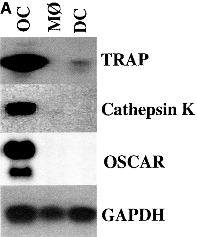
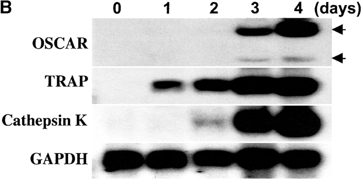
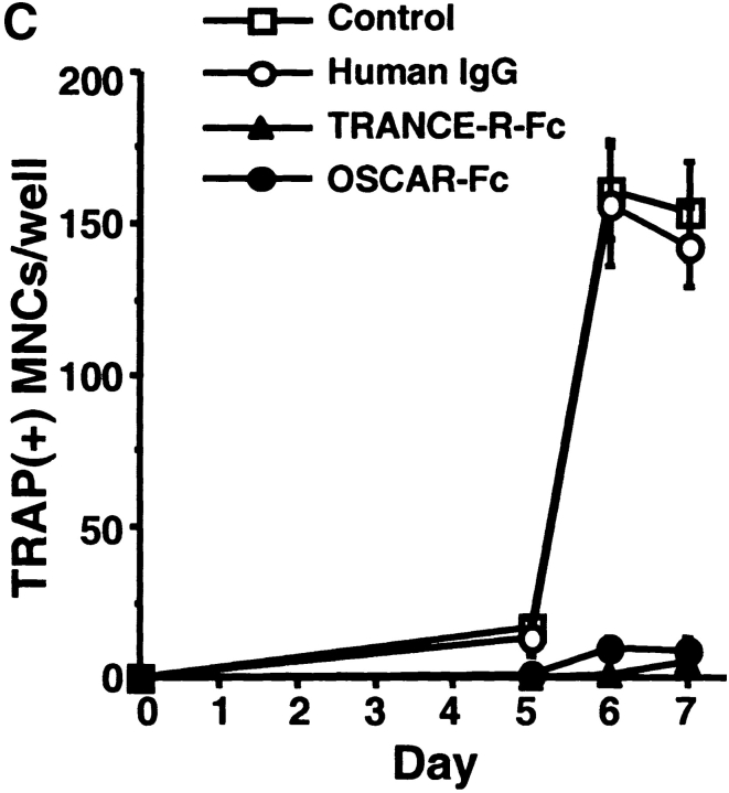
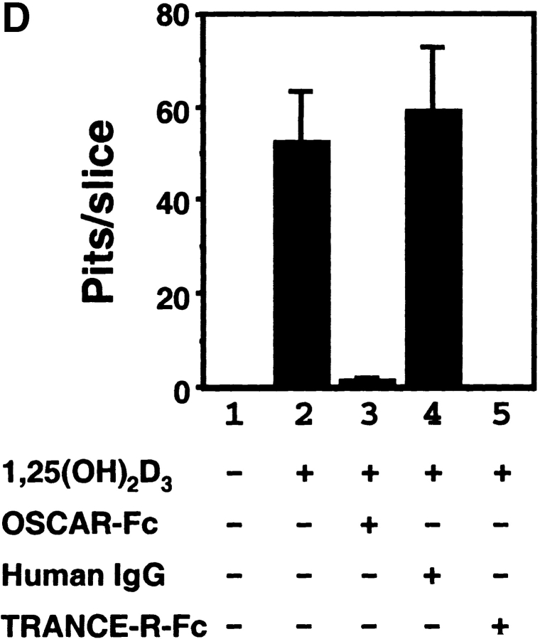
Osteoclasts (OCs) are multinucleated cells that resorb bone and are essential for bone homeostasis. They develop from hematopoietic cells of the myelomonocytic lineage. OC formation requires cell-to-cell interactions with osteoblasts and can be achieved by coculturing bone marrow precursor cells with osteoblasts/stromal cells. Two of the key factors mediating the osteoblast-induced osteoclastogenesis are macrophage-colony stimulating factor (M-CSF) and the tumor necrosis factor (TNF) family member TNF-related activation-induced cytokine (TRANCE) that are produced by osteoblasts/stromal cells in response to various bone resorbing hormones. In addition, other factors produced by osteoblasts/stromal cells further influence osteoclastogenesis. Here we report the identification and characterization of OC-associated receptor (OSCAR), a novel member of the leukocyte receptor complex (LRC)-encoded family expressed specifically in OCs. Genes in the LRC produce immunoglobulin (Ig)-like surface receptors and play critical roles in the regulation of both innate and adaptive immune responses. Different from the previously characterized members of the LRC complex, OSCAR expression is detected specifically in preosteoclasts or mature OCs. Its putative-ligand (OSCAR-L) is expressed primarily in osteoblasts/stromal cells. Moreover, addition of a soluble form of OSCAR in coculture with osteoblasts inhibits the formation of OCs from bone marrow precursor cells in the presence of bone-resorbing factors, indicating that OSCAR may be an important bone-specific regulator of OC differentiation. In addition, this study suggests that LRC-encoded genes may have evolved to regulate the physiology of cells beyond those of the immune system.


Figures

Figure 1.
Figure 1.
OSCAR mRNA expression. (A) Northern blot analysis of RNA from bone marrow–derived OCs OC, macrophages (MØ), and DCs. GAPDH was used as control. (B) OSCAR mRNA expressions during the differentiation of RAW264.7 to mature OCs by TRANCE. Northern blot analysis was performed using total RNA from RAW264.7 cells stimulated with recombinant TRANCE (200 ng/ml) for 0–4 d. OSCAR, TRAP, Cathepsin K, and GAPDH were used for probes. (C and D) Northern blot analysis of OSCAR in various mouse tissues and bone marrow–derived OCs.
Figure 1.
Figure 1.
OSCAR mRNA expression. (A) Northern blot analysis of RNA from bone marrow–derived OCs OC, macrophages (MØ), and DCs. GAPDH was used as control. (B) OSCAR mRNA expressions during the differentiation of RAW264.7 to mature OCs by TRANCE. Northern blot analysis was performed using total RNA from RAW264.7 cells stimulated with recombinant TRANCE (200 ng/ml) for 0–4 d. OSCAR, TRAP, Cathepsin K, and GAPDH were used for probes. (C and D) Northern blot analysis of OSCAR in various mouse tissues and bone marrow–derived OCs.
Figure 2.
Figure 2.
Sequence analysis of OSCAR. (A) The predicted amino acid sequence of the full-length murine OSCAR protein (mOSCAR) compared with that of human OSCAR (hOSCAR). Dots indicate shared identities between mouse and human protein, and dashes indicate gaps between regions of homology. The boldface letters indicate the position of the conserved cysteine in the Ig-fold. Residues labeled with an asterisk (*) indicate potential NH2-linked glycosylation sites. The numbers in left-hand column indicate the amino acid residue positions in the mOSCAR and hOSCAR protein. Between L12 and W13 of mOSCAR, six amino acids(CELSLP) are inserted by alternative splicing in the minor population of mOSCAR cDNA clones. Also, 1 of 15 clones of hOSCAR has a four amino acid (AIIV) insertion between V24 and P25 of hOSCAR and 2 of 15 clones of hOSCAR have a deletion of 11 amino acids (residues 13 through 24, WPLCHTDITPSV, are replaced with F13) by alternative splicing. GenBank accession nos. for mOSCAR are AF391159, AF391160, and AF391161 and for hOSCAR are AF391162, AF391163, and AF391164. (B) The sequence comparison of the two Ig-like domains of hOSCAR with other members of the LRC-encoded genes: hIGSF(AF034198), hKIR(AF072410), hILT1(U82275), and hGPVI(AB035073). Gaps in alignment are indicated by dashes and identical amino acids appear on a dark background. The numbers in the left-hand column indicate the residue positions from the full-length protein sequences.
Figure 2.
Figure 2.
Sequence analysis of OSCAR. (A) The predicted amino acid sequence of the full-length murine OSCAR protein (mOSCAR) compared with that of human OSCAR (hOSCAR). Dots indicate shared identities between mouse and human protein, and dashes indicate gaps between regions of homology. The boldface letters indicate the position of the conserved cysteine in the Ig-fold. Residues labeled with an asterisk (*) indicate potential NH2-linked glycosylation sites. The numbers in left-hand column indicate the amino acid residue positions in the mOSCAR and hOSCAR protein. Between L12 and W13 of mOSCAR, six amino acids(CELSLP) are inserted by alternative splicing in the minor population of mOSCAR cDNA clones. Also, 1 of 15 clones of hOSCAR has a four amino acid (AIIV) insertion between V24 and P25 of hOSCAR and 2 of 15 clones of hOSCAR have a deletion of 11 amino acids (residues 13 through 24, WPLCHTDITPSV, are replaced with F13) by alternative splicing. GenBank accession nos. for mOSCAR are AF391159, AF391160, and AF391161 and for hOSCAR are AF391162, AF391163, and AF391164. (B) The sequence comparison of the two Ig-like domains of hOSCAR with other members of the LRC-encoded genes: hIGSF(AF034198), hKIR(AF072410), hILT1(U82275), and hGPVI(AB035073). Gaps in alignment are indicated by dashes and identical amino acids appear on a dark background. The numbers in the left-hand column indicate the residue positions from the full-length protein sequences.
Figure 3.
Figure 3.
Expression and regulation of OSCAR protein. (A) FACS® analysis of cell surface OSCAR expression on freshly isolated bone marrow cells (BM), bone marrow–derived MØ (BM-MØ), and OCs (BM-OC). After differentiation, cells were stained with anti-OSCAR mAb (open histogram) or isotype control mAb (shaded histogram). (B) Immunohistochemistry of OSCAR on in vitro bone marrow–derived macrophage (BM-MØ) and bone marrow–derived OC (BM-OC). For immunohistochemistry, cells were fixed with 4% paraformaldehyde, and followed by TRAP assay (left and middle), staining with rat anti-OSCAR mAb (middle and right) or rat IgG as a control (left). TRAP-positive cells are stained red and OSCAR-expressing cells are stained blue.
Figure 4.
Figure 4.
Role of OSCAR in OC differentiation. (A) A soluble form of OSCAR inhibits OC formation from bone marrow precursor cells in the coculture with osteoblast/stromal cells in the presence of osteotropic factors. Mouse bone marrow cells (105 cells/ per well) collected from 5–7-wk-old male mice and mouse primary calvarial osteoblasts (104 cells per well) collected from newborn mice were cocultured in the presence of 10−8 M 1α,25-dihydroxyvitamin D3 (1,25(OH)2D3) with 20 μg/ml OSCAR-Fc, 20 μg/ml human IgG, or 5 μg/ml TRANCE-R-Fc in 96-well culture plates (top) or on dentine slices (4 mm diameter) placed in 96-well culture plates (bottom). Cells were also cultured in the presence of 10−8 M 1,25(OH)2D3 without any additives as a control. On day 6, cells in 96-well culture plates were fixed and stained for TRAP. TRAP-positive cells appeared as red cells (top). Cells on dentine slices were removed, and dentine slices were stained with Mayer's hematoxylin. Pits appeared as dark spots (bottom). Bar indicates 250 μm. In the absence of 1,25(OH)2D3, no TRAP-positive multinucleated cells appeared in the coculture (data not shown). (B) Mouse bone marrow cells and primary calvarial osteoblasts were cocultured in the presence of 10−8 M 1,25(OH)2D3 with increasing concentrations (0, 5, 10, and 20 μg/ml) of OSCAR–Fc (black circles) or human IgG (white circles) in 96-well culture plates for 6 d. After fixing and staining cells for TRAP, numbers of TRAP-positive multinucleated cells (MNCs) were counted as OCs. (C) Mouse bone marrow cells and mouse primary calvarial osteoblasts were cocultured in the presence of 10−8 M 1,25(OH)2D3 with 20 μg/ml OSCAR–Fc (black circles), 20 μg/ml human IgG (white circles), or 5μg/ml TRANCE-R-Fc (black triangles) in 96-well culture plates. Cells were also cultured in the presence of 10−8 M 1,25(OH)2D3 without any additives as control (white squares). On days 0, 5, 6, and 7, cells were fixed and stained for TRAP. TRAP-positive multinucleated cells were counted as OCs. (D) Number of pits formed on dentine slices in A was counted. Media of each culture were replaced with fresh media on day 3 (A–D). Results are representative of at least three independent sets of similar experiments (A–D). All data are expressed as mean ± SD of quadruplicate cultures.
Figure 4.
Figure 4.
Role of OSCAR in OC differentiation. (A) A soluble form of OSCAR inhibits OC formation from bone marrow precursor cells in the coculture with osteoblast/stromal cells in the presence of osteotropic factors. Mouse bone marrow cells (105 cells/ per well) collected from 5–7-wk-old male mice and mouse primary calvarial osteoblasts (104 cells per well) collected from newborn mice were cocultured in the presence of 10−8 M 1α,25-dihydroxyvitamin D3 (1,25(OH)2D3) with 20 μg/ml OSCAR-Fc, 20 μg/ml human IgG, or 5 μg/ml TRANCE-R-Fc in 96-well culture plates (top) or on dentine slices (4 mm diameter) placed in 96-well culture plates (bottom). Cells were also cultured in the presence of 10−8 M 1,25(OH)2D3 without any additives as a control. On day 6, cells in 96-well culture plates were fixed and stained for TRAP. TRAP-positive cells appeared as red cells (top). Cells on dentine slices were removed, and dentine slices were stained with Mayer's hematoxylin. Pits appeared as dark spots (bottom). Bar indicates 250 μm. In the absence of 1,25(OH)2D3, no TRAP-positive multinucleated cells appeared in the coculture (data not shown). (B) Mouse bone marrow cells and primary calvarial osteoblasts were cocultured in the presence of 10−8 M 1,25(OH)2D3 with increasing concentrations (0, 5, 10, and 20 μg/ml) of OSCAR–Fc (black circles) or human IgG (white circles) in 96-well culture plates for 6 d. After fixing and staining cells for TRAP, numbers of TRAP-positive multinucleated cells (MNCs) were counted as OCs. (C) Mouse bone marrow cells and mouse primary calvarial osteoblasts were cocultured in the presence of 10−8 M 1,25(OH)2D3 with 20 μg/ml OSCAR–Fc (black circles), 20 μg/ml human IgG (white circles), or 5μg/ml TRANCE-R-Fc (black triangles) in 96-well culture plates. Cells were also cultured in the presence of 10−8 M 1,25(OH)2D3 without any additives as control (white squares). On days 0, 5, 6, and 7, cells were fixed and stained for TRAP. TRAP-positive multinucleated cells were counted as OCs. (D) Number of pits formed on dentine slices in A was counted. Media of each culture were replaced with fresh media on day 3 (A–D). Results are representative of at least three independent sets of similar experiments (A–D). All data are expressed as mean ± SD of quadruplicate cultures.
Figure 5.
Figure 5.
Expression of OSCAR-L in osteoblasts. Primary calvarial osteoblasts are derived as described in the Materials and Methods. Surface OSCAR-L expression on various cells was determined by FACS® analysis using control human IgG1 (dotted lines) or OSCAR-Fc (solid lines) and FITC-labeled anti–human IgG1. A thymoma cell line, BW5147, which does not express OSCAR-L, was also shown here.

References

    1. Suda, T., N. Takahashi, N. Udagawa, E. Jimi, M.T. Gillespie, and T.J. Martin. 1999. Modulation of osteoclast differentiation and function by the new members of the tumor necrosis factor receptor and ligand families. Endocr. Rev. 20:345–357. - PubMed
    1. Teitelbaum, S.L. 2000. Bone resorption by osteoclasts. Science. 289:1504–1508. - PubMed
    1. Wiktor-Jedrzejczak, W., A. Bartocci, A.W. Ferrante, Jr., A. Ahmed-Ansari, K.W. Sell, J.W. Pollard, and E.R. Stanley. 1990. Total absence of colony-stimulating factor 1 in the macrophage-deficient osteopetrotic (op/op) mouse. Proc. Natl. Acad. Sci. USA. 87:4828–4832. - PMC - PubMed
    1. Yoshida, H., S. Hayashi, T. Kunisada, M. Ogawa, S. Nishikawa, H. Okamura, T. Sudo, and L.D. Shultz. 1990. The murine mutation osteopetrosis is in the coding region of the macrophage colony stimulating factor gene. Nature. 345:442–444. - PubMed
    1. Yasuda, H., N. Shima, N. Nakagawa, K. Yamaguchi, M. Kinosaki, S. Mochizuki, A. Tomoyasu, K. Yano, M. Goto, A. Murakami, et al. 1998. Osteoclast differentiation factor is a ligand for osteoprotegerin/osteoclastogenesis-inhibitory factor and is identical to TRANCE/RANKL. Proc. Natl. Acad. Sci. USA. 95:3597–3602. - PMC - PubMed

Publication types