. 2002 Jan 21;195(2):211-20.
doi: 10.1084/jem.200116116.

Complement C3 activation is required for antiphospholipid antibody-induced fetal loss

Affiliations

Complement C3 activation is required for antiphospholipid antibody-induced fetal loss

V Michael Holers et al. J Exp Med. .

Abstract

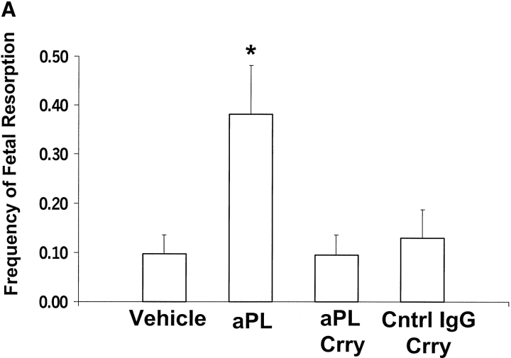
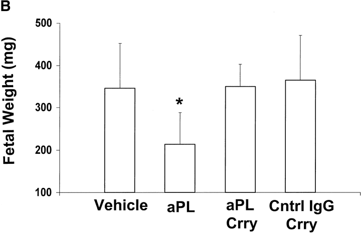
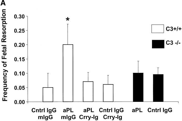
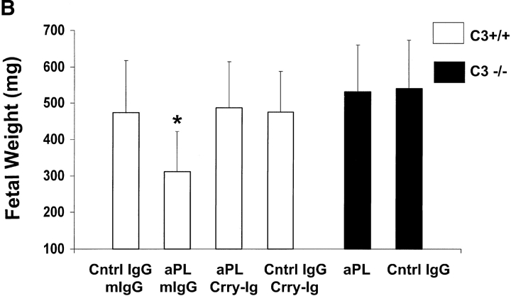
The antiphospholipid syndrome (APS) is characterized by recurrent fetal loss, vascular thrombosis, and thrombocytopenia occurring in the presence of antiphospholipid (aPL) antibodies. The pathogenesis of fetal loss and tissue injury in APS is incompletely understood, but is thought to involve platelet and endothelial cell activation as well as procoagulant effects of aPL antibodies acting directly on clotting pathway components. Recent studies have shown that uncontrolled complement activation in the placenta leads to fetal death in utero. We hypothesized that aPL antibodies activate complement in the placenta, generating split products that mediate placental injury and lead to fetal loss and growth retardation. To test this hypothesis, we used a murine model of APS in which pregnant mice are injected with human IgG containing aPL antibodies. We found that inhibition of the complement cascade in vivo, using the C3 convertase inhibitor complement receptor 1-related gene/protein y (Crry)-Ig, blocks fetal loss and growth retardation. Furthermore, mice deficient in complement C3 were resistant to fetal injury induced by aPL antibodies. While antigenic epitopes recognized by aPL antibodies are important in the pathogenesis of APS, our data show that in vivo complement activation is required for aPL antibody-induced fetal loss and growth retardation.


Figures

Figure 2.
Figure 2.
Effects of Crry-Ig on aPL antibody-induced fetal resorption. Representative uteri from BALB/c mice killed at day 15 of pregnancy are shown. The top panel, from a mouse treated with aPL-IgG and murine IgG, shows eight fetuses and deciduas of varying size and two resorptions (arrows). The bottom panel, from a mouse treated with aPL and Crry-Ig, contains larger fetuses with deciduas and no resorptions.
Figure 1.
Figure 1.
Crry-Ig ameliorates aPL-IgG–induced pregnancy complications. Female BALB/c mice were treated intraperitoneally with IgG (10 mg) from a patient with APS (aPL), normal human IgG (Cntrl IgG), or saline (Vehicle) on days 8 and 12 of pregnancy. Some the mice received Crry-Ig (3 mg intraperitoneal) every other day from days 8–12. Mice were killed on day 15 of pregnancy, uteri were dissected, fetuses were weighed, and frequency of fetal resorption calculated (number of resorptions/number of fetuses plus number of resorptions). There were six mice in each group. (A) Treatment with aPL-IgG caused an increase in fetal resorptions compared with vehicle or control human IgG (*P < 0.05) which was prevented by Crry-Ig (*aPL versus aPL plus Crry-Ig; P < 0.05). (B) aPL-IgG caused fetal growth retardation (*aPL versus Cntrl IgG; P < 0.01) which was also prevented by Crry-Ig (*aPL versus aPL plus Crry-Ig; P < 0.01).
Figure 1.
Figure 1.
Crry-Ig ameliorates aPL-IgG–induced pregnancy complications. Female BALB/c mice were treated intraperitoneally with IgG (10 mg) from a patient with APS (aPL), normal human IgG (Cntrl IgG), or saline (Vehicle) on days 8 and 12 of pregnancy. Some the mice received Crry-Ig (3 mg intraperitoneal) every other day from days 8–12. Mice were killed on day 15 of pregnancy, uteri were dissected, fetuses were weighed, and frequency of fetal resorption calculated (number of resorptions/number of fetuses plus number of resorptions). There were six mice in each group. (A) Treatment with aPL-IgG caused an increase in fetal resorptions compared with vehicle or control human IgG (*P < 0.05) which was prevented by Crry-Ig (*aPL versus aPL plus Crry-Ig; P < 0.05). (B) aPL-IgG caused fetal growth retardation (*aPL versus Cntrl IgG; P < 0.01) which was also prevented by Crry-Ig (*aPL versus aPL plus Crry-Ig; P < 0.01).
Figure 3.
Figure 3.
APL antibody-induced necrosis and infiltration of neutrophils in decidual tissue is prevented by Crry-Ig. Pregnant mice (day 8) were injected with control human IgG (A and D), aPL-IgG (B and E), or aPL-IgG and Crry-Ig (C and F), and uteri were removed after 90 min. In mice treated with aPL-IgG there was increased density of cells and focal necrosis within the decidual tissue (B) and clumping, apoptosis, and scattered clusters of neutrophils (arrows) throughout (E). Decidual tissue from mice treated with aPL-IgG and Crry-Ig was similar to mice treated with control Ig, showing uniform, sparse cellularity (A and C) and normal appearing trophoblastic cells (D and F). Sections were stained with H&E. (A–C) Original magnification: ×10; (D–F) original magnification: ×40.
Figure 4.
Figure 4.
Complement activation in decidual tissue of aPL antibody-treated mice is prevented by Crry-Ig. Sections of decidual tissue from mice injected with control human IgG (A), aPL-IgG (B), or aPL-IgG and Crry-Ig (C) as described in Fig. 3 were stained with antibody to mouse C3. In the aPL-IgG treated mice (B), there was extensive C3 deposition in the decidua and extraembryonic membranes. In contrast, the decidual tissues from mice treated with control IgG (A) or aPL-IgG and Crry-Ig (C) show minimal staining for C3 with more intense staining on the extraembryonic tissues (arrows). Original magnification: ×10.
Figure 5.
Figure 5.
C3-deficient mice are protected from aPL antibody-induced pregnancy complications. C3+/+ mice (B6/Sv129F1) were treated with aPL-IgG (10 mg intraperitoneally) (aPL) or normal human IgG (Cntrl IgG) on days 8 and 12 of pregnancy. Half of the mice in each group received Crry-Ig (3 mg intraperitoneally) every other day from days 8–12 and half received control murine IgG (mIgG). C3−/− mice were treated with either aPL-IgG or normal human IgG. Pregnancy outcomes were assessed as described in the legend for Fig. 1. There were 10–14 mice in each experimental group. (A) Analysis of the four groups of C3+/+ mice shows that treatment with aPL-IgG caused an increase in frequency of fetal resorptions in this strain (*aPL plus mIgG versus Cntrl IgG plus mIgG; P < 0.01), while C3−/− were protected from aPL-induced pregnancy loss (aPL versus Cntrl IgG; P = NS). In the C3+/+ mice, Crry-Ig prevented aPL-induced fetal resorption (*aPL plus mIgG versus aPL plus Crry-Ig; P < 0.01). (B) Similarly, aPL treatment caused a decrease in fetal weight in C3+/+ mice (*aPL plus mIgG versus Cntrl IgG plus mIgG; P < 0.01) which was absent in C3−/− mice, and this was ameliorated by Crry-Ig (*aPL plus mIgG versus aPL plus Crry-Ig; P < 0.01).
Figure 5.
Figure 5.
C3-deficient mice are protected from aPL antibody-induced pregnancy complications. C3+/+ mice (B6/Sv129F1) were treated with aPL-IgG (10 mg intraperitoneally) (aPL) or normal human IgG (Cntrl IgG) on days 8 and 12 of pregnancy. Half of the mice in each group received Crry-Ig (3 mg intraperitoneally) every other day from days 8–12 and half received control murine IgG (mIgG). C3−/− mice were treated with either aPL-IgG or normal human IgG. Pregnancy outcomes were assessed as described in the legend for Fig. 1. There were 10–14 mice in each experimental group. (A) Analysis of the four groups of C3+/+ mice shows that treatment with aPL-IgG caused an increase in frequency of fetal resorptions in this strain (*aPL plus mIgG versus Cntrl IgG plus mIgG; P < 0.01), while C3−/− were protected from aPL-induced pregnancy loss (aPL versus Cntrl IgG; P = NS). In the C3+/+ mice, Crry-Ig prevented aPL-induced fetal resorption (*aPL plus mIgG versus aPL plus Crry-Ig; P < 0.01). (B) Similarly, aPL treatment caused a decrease in fetal weight in C3+/+ mice (*aPL plus mIgG versus Cntrl IgG plus mIgG; P < 0.01) which was absent in C3−/− mice, and this was ameliorated by Crry-Ig (*aPL plus mIgG versus aPL plus Crry-Ig; P < 0.01).
Figure 6.
Figure 6.
aPL-IgG–induced thrombophilia is inhibited by Crry-Ig. CD-1 mice were injected intraperitoneal with affinity purified aPL-IgG (aPL) or normal human (Cntrl IgG) at 0 h and 48 h. Half the mice in each group received Crry-Ig, and half the mice received control murine IgG (mIgG). At 72 h after the first injection, surgically induced thrombus formation was measured as described in Materials and Methods. There were 11–14 mice in each experimental group. Treatment with aPL-IgG caused an increase in thrombus size (*aPL plus mIgG versus Cntrl IgG plus mIgG; P < 0.05), while Crry-Ig prevented aPL-induced enhancement of thrombosis (*aPL plus mIgG versus aPL plus Crry-Ig; P < 0.05; Cntrl IgG plus Crry-Ig versus aPL plus Crry-Ig; P = NS).

Similar articles

Cited by

References

    1. Branch, D.W., D.J. Dudley, M.D. Mitchell, K.A. Creighton, T.M. Abbott, E.H. Hammond, and R.A. Daynes. 1990. Immunoglobulin G fractions from patients with antiphospholipid antibodies cause fetal death in BALB/c mice: a model for autoimmune fetal loss. Am. J. Obstet. Gynecol. 163:210–216. - PubMed
    1. Piona, A., L. La Rosa, A. Tincani, D. Faden, G. Magro, S. Grasso, F. Nicoletti, G. Balestrieri, and P.L. Meroni. 1995. Placental thrombosis and fetal loss after passive transfer of mouse lupus monoclonal or human polyclonal anti-cardiolipin antibodies in pregnant naive BALB/c mice. Scand. J. Immunol. 41:427–432. - PubMed
    1. Blank, M., J. Cohen, V. Toder, and Y. Shoenfeld. 1991. Induction of antiphospholipid syndrome in naive mice with mouse lupus monoclonal and human polyclonal antibodies. Proc. Natl. Acad. Sci. USA. 88:3069–3073. - PMC - PubMed
    1. Ikematsu, W., F.L. Luan, L. La Rosa, B. Beltrami, F. Nicoletti, J.P. Buyon, P.L. Meroni, G. Balestrieri, and P. Casali. 1998. Human anticardiolipin monoclonal autoantibodies cause placental necrosis and fetal loss in BALB/c mice. Arthr. Rheum. 41:1026–1039. - PubMed
    1. Di Simone, N., P.L. Meroni, N. Del Papa, E. Raschi, D. Caliandro, S. del Carlolis, M.A. Khamashta, T. Atsuni, G.R.V. Hughes, G. Balestrieri, et al. 2000. Antiphospholipid antibodies affect trophoblast gonadotropin secretion and invasiveness by binding directly and through adhered B2-glycoprotein 1. Arthr. Rheum. 43:140–150. - PMC - PubMed

Publication types