. 2002 Jan 21;195(2):233-44.
doi: 10.1084/jem.20011885.

Requirement for the NF-kappaB family member RelA in the development of secondary lymphoid organs

Affiliations

Requirement for the NF-kappaB family member RelA in the development of secondary lymphoid organs

Elizabeth Alcamo et al. J Exp Med. .

Abstract

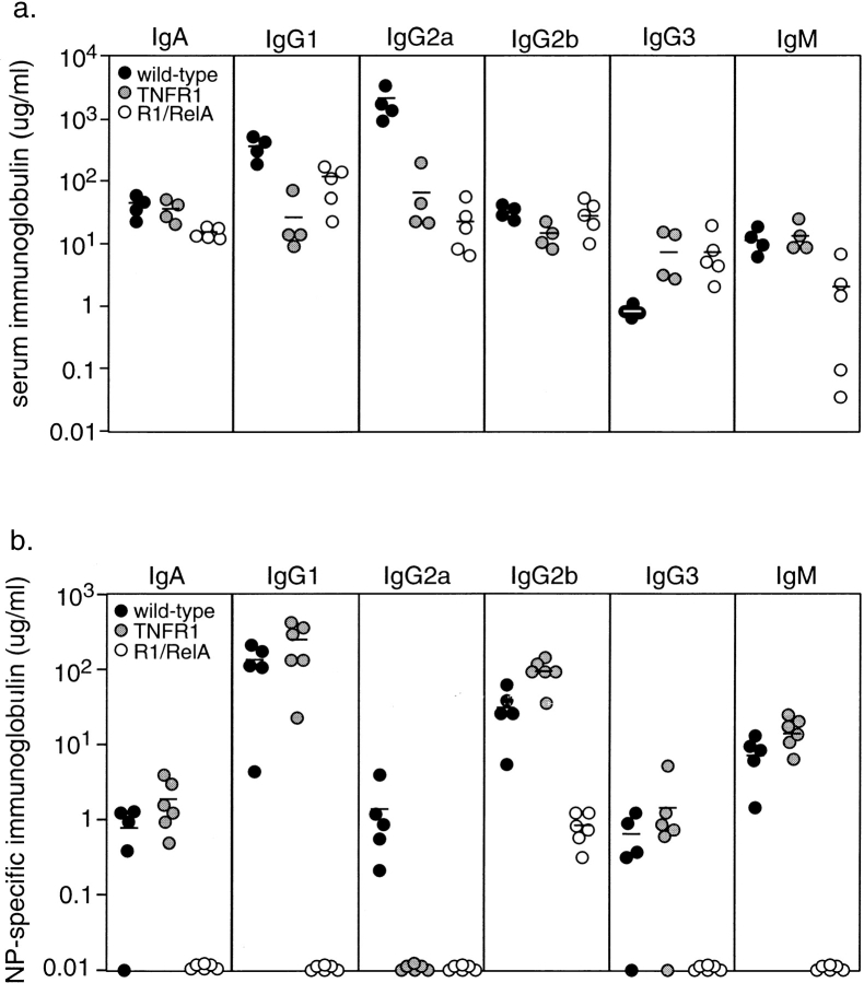
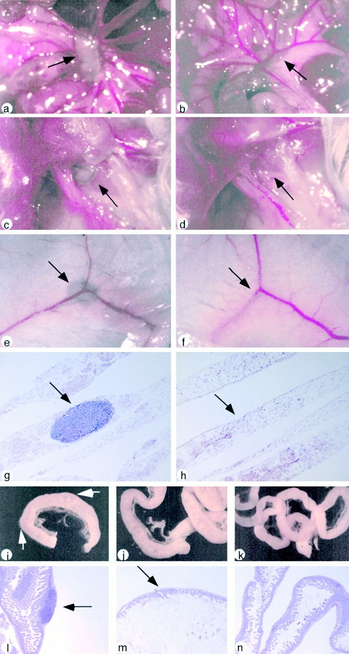
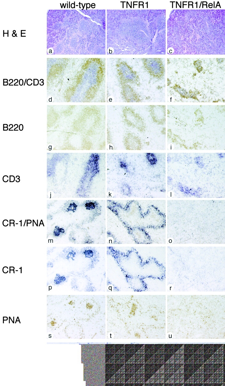
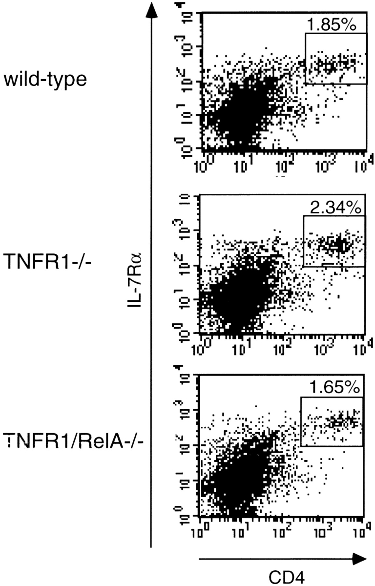
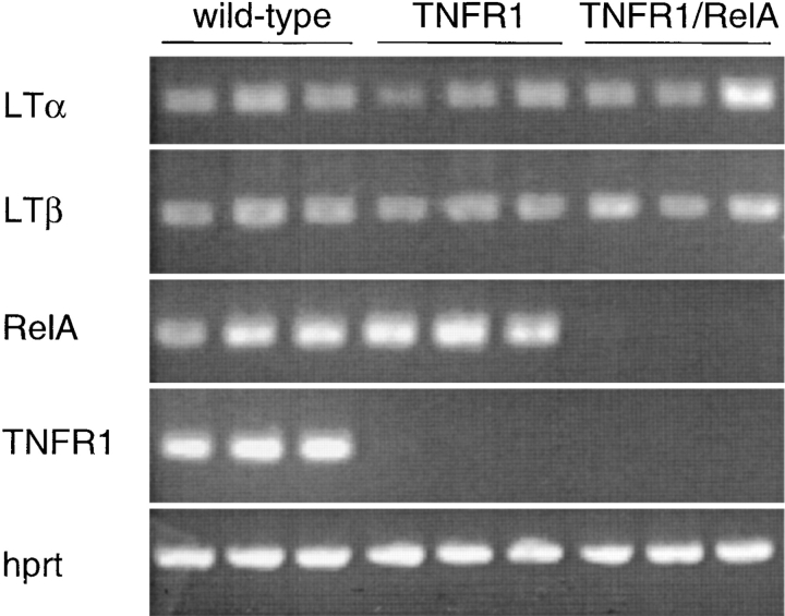
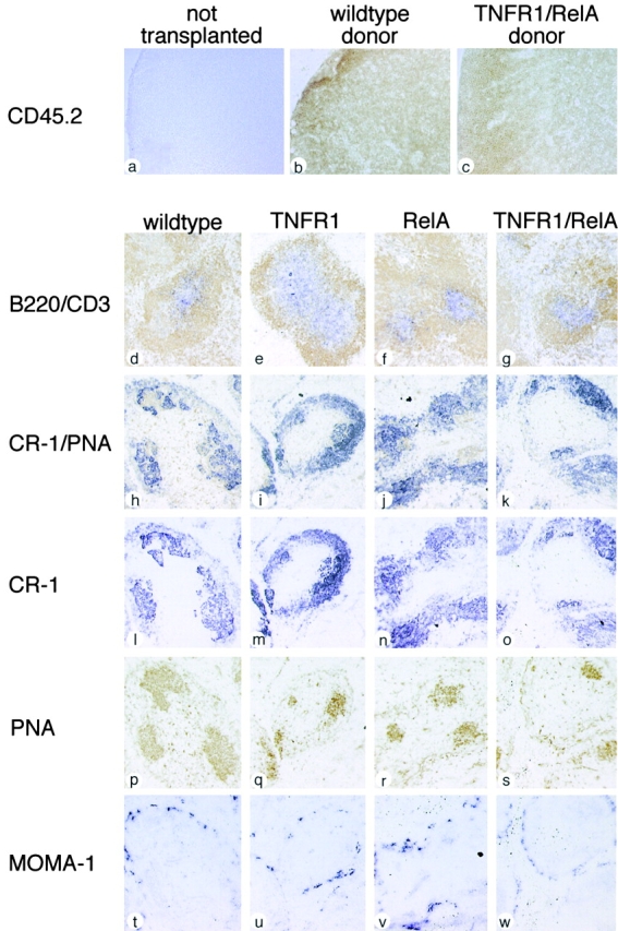
The transcription factor nuclear factor (NF)-kappaB has been suggested to be a key mediator of the development of lymph nodes and Peyer's patches. However, targeted deletion of NF-kappaB/ Rel family members has not yet corroborated such a function. Here we report that when mice lacking the RelA subunit of NF-kappaB are brought to term by breeding onto a tumor necrosis factor receptor (TNFR)1-deficient background, the mice that are born lack lymph nodes, Peyer's patches, and an organized splenic microarchitecture, and have a profound defect in T cell-dependent antigen responses. Analyses of TNFR1/RelA-deficient embryonic tissues and of radiation chimeras suggest that the dependence on RelA is manifest not in hematopoietic cells but rather in radioresistant stromal cells needed for the development of secondary lymphoid organs.


Figures

Figure 1.
Figure 1.
Severely reduced T cell–dependent antibody response in TNFR1/RelA-deficient mice in vivo. (a) Basal immunoglobulin concentrations. Serum was collected from 3-wk-old wild-type (black circle), TNFR1-deficient (gray circle), and TNFR1/RelA-deficient (open circle) mice and immunoglobulin concentrations determined by isotype-specific ELISA. (b) T cell–dependent antibody production. 3-wk-old mice were immunized with NP-CG adsorbed to alum and the level of NP-specific antibodies determined 1 wk later by ELISA using NP-BSA as capture antibody. Each point represents the mean of duplicate assays from one animal; horizontal bars indicate mean concentration of the group.
Figure 2.
Figure 2.
Absence of LNs and PPs in TNFR1/RelA-deficient mice. Five 3-wk-old wild-type (a, c, e, g, i, and l), TNFR1-deficient (j and m) and TNFR1/RelA-deficient (b, d, f, h, k, and n) mice were injected intraperitoneally with Evans Blue (1 mg/ml, 20 μl per mouse) 18 h before dissection to visualize LNs and PPs. (a and b) mesenteries; (c and d) lateral axillary fossae; (e and f) inguinal fat pad; (g and h) inguinal fat pad, sectioned and H & E-stained; (i, j, and k) intestine whole mounts; (l, m, and n) intestine, sectioned and H & E-stained. Arrows indicate regions in which LNs and PPs should be found. 15 additional mice of each mutant genotype were inspected for the presence of these organs without prior injection of Evan Blue; similar results were observed.
Figure 3.
Figure 3.
Aberrant organization of lymphocytes and absence of secondary immune response structures in TNFR1/RelA-deficient spleens. 3-wk-old wild-type (a, d, g, j, m, p, s, and v), TNFR1-deficient (b, e, h, k, n, q, t, and w), and TNFR1/RelA-deficient mice (c, f, i, l, o, r, u, and x) were immunized with NP7-CG and killed 1 wk later for analysis of splenic structures. Two wild-type, two TNFR1-deficient, and five TNFR1/RelA-deficient spleens were assayed. Sequential sections of spleen were stained with (a–c) H & E; (d–l) anti-B220 (brown) and/or anti-CD3 (purple); (m–u) anti-CR-1 (purple) and/or PNA (brown); and (v–x) MOMA-1 (purple).
Figure 4.
Figure 4.
Presence of CD4+CD3IL-7Rα+ cells in TNFR1/RelA-deficient embryonic intestine. Flow cytometry was used to analyze intestinal cells from E17.5 wild-type, TNFR1-deficient, and TNFR1/RelA fetuses with the indicated cell-surface markers. All events presented are negative for CD3. Each sample assayed consisted of tissue from three embryos of the same genotype. Similar results were obtained from two independent FACS® experiments.
Figure 5.
Figure 5.
Normal expression of LTα1β2 in TNFR1/RelA-deficient embryonic intestine. RNA was prepared from E17.5 wild-type, TNFR1-deficient, TNFR1/RelA-deficient intestines and assayed by RT-PCR for expression of LTα and LTβ, RelA, TNFR1, and hprt (control).
Figure 6.
Figure 6.
Repopulation of LNs and organization of spleen in adoptive transfer mice reconstituted with TNFR1/RelA-deficient hematopoietic cells. C57BL/6 CD45.1+ mice were lethally irradiated and transplanted with wild-type, TNFR1-deficient, RelA-deficient, or TNFR1/RelA-deficient fetal liver cells. 4 mo after transplant, animals were killed and tissues removed for cryosectioning. LNs from one animal and spleens from two animals of each genotype were assayed. (a–c) Inguinal LNs stained with donor-specific anti-CD45.2 (brown). (d–w) Sequential sections of spleen stained with (d–g) anti-B220 (brown) and/or anti-CD3 (purple); (h–s) anti-CR-1 (purple) and/or PNA (brown); and (t–w) anti-MOMA-1 (purple).

Similar articles

Cited by

References

    1. Karrer, U., A. Althage, B. Odermatt, C.W. Roberts, S.J. Korsmeyer, S. Miyawaki, H. Hengartner, and R.M. Zinkernagel. 1997. On the key role of secondary lymphoid organs in antiviral immune responses studied in alymphoplastic (aly/aly) and spleenless (Hox11(−)/−) mutant mice. J. Exp. Med. 185:2157–2170. - PMC - PubMed
    1. Shinkura, R., K. Kitada, F. Matsuda, K. Tashiro, K. Ikuta, M. Suzuki, K. Kogishi, T. Serikawa, and T. Honjo. 1999. Alymphoplasia is caused by a point mutation in the mouse gene encoding Nf-kappa b-inducing kinase. Nat. Genet. 22:74–77. - PubMed
    1. Dougall, W.C., M. Glaccum, K. Charrier, K. Rohrbach, K. Brasel, T. De Smedt, E. Daro, J. Smith, M.E. Tometsko, C.R. Maliszewski, et al. 1999. RANK is essential for osteoclast and lymph node development. Genes Dev. 13:2412–2424. - PMC - PubMed
    1. Kong, Y.Y., H. Yoshida, I. Sarosi, H.L. Tan, E. Timms, C. Capparelli, S. Morony, A.J. Oliveira-dos-Santos, G. Van, A. Itie, et al. 1999. OPGL is a key regulator of osteoclastogenesis, lymphocyte development and lymph-node organogenesis. Nature. 397:315–323. - PubMed
    1. Kim, D., R.E. Mebius, J.D. MacMicking, S. Jung, T. Cupedo, Y. Castellanos, J. Rho, B.R. Wong, R. Josien, N. Kim, et al. 2000. Regulation of peripheral lymph node genesis by the tumor necrosis factor family member TRANCE. J. Exp. Med. 192:1467–1478. - PMC - PubMed

Publication types