. 2002 Feb 5;99(3):1235-40.
doi: 10.1073/pnas.022631899. Epub 2002 Jan 22.

Subnuclear distribution of topoisomerase I is linked to ongoing transcription and p53 status

Affiliations

Subnuclear distribution of topoisomerase I is linked to ongoing transcription and p53 status

Yinghui Mao et al. Proc Natl Acad Sci U S A. .

Erratum in

  • Proc Natl Acad Sci U S A 2002 Mar 19;99(6):4134

Abstract

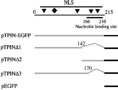
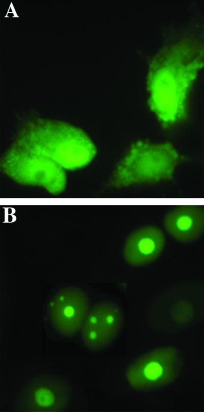
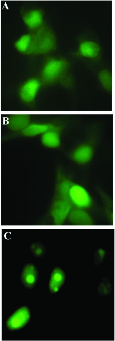
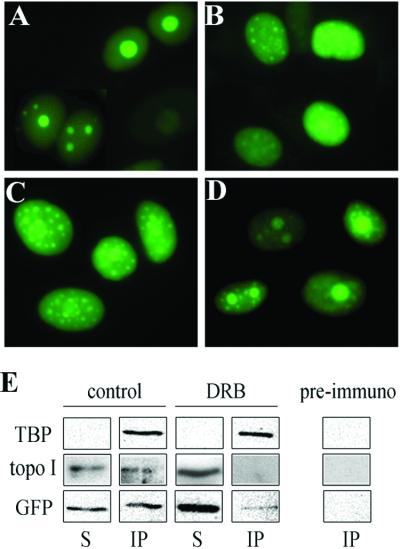
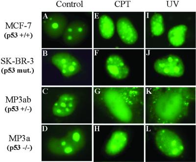
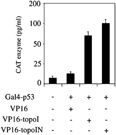
The nonconserved, hydrophilic N-terminal domain of eukaryotic DNA topoisomerase I (topo I) is dispensable for catalytic activity in vitro but essential in vivo. There are at least five putative nuclear localization signals and a nucleolin-binding signal within the first 215 residues of the topo I N-terminal domain. We have investigated physiological functions of the topo I N-terminal domain by fusing it to an enhanced green fluorescent protein (EGFP). The first 170 residues of the N-terminal domain allow efficient import of chimeric proteins into nuclei and nucleoli. The nucleolar localization of this protein does not depend on its interaction with nucleolin, whereas ongoing rDNA transcription clearly is crucial. Immunoprecipitation experiments reveal that the topo I N terminus (topoIN)-EGFP fusion protein associates with the TATA-binding protein in cells. Furthermore, DNA damage results in extensive nuclear redistribution of the topoIN-EGFP chimeric product. The redistribution is also p53-dependent and the N terminus of topo I appears to interact with p53 in vivo. These results show that the topo I localization to the nucleolus is related to the p53 and DNA damage, as well as changes in transcriptional status. Nucleolar release of topo I under conditions of cellular duress may represent an important, antecedent step in tumor cell killing by topoisomerase active agents.


Figures

Figure 1
Figure 1
Schematic description of topoIN-EGFP constructs. The N-terminal domain of topo I and its deleted fragments were cloned into pEGFPN2. The top line shows the topo I N terminus (residues 1–215). Four putative NLSs (triangles) are located at residues 59–65 (KKHKEKE), residues 150–156 (KKIKTED), residues 174–180 (KKPKNKED), and residues 192–198 (KKKPKKE) (18). A novel NLS is marked by the diamond at residues 117–146 (DEDDAD or KDEPEDDG) (20). The nucleolin-binding region is located from residues 166 to 210 as shown. The lower collection of lines represent fusion constructs of N-terminal segments of topo I (light line) and EGFP (heavy line), and numbers correspond to amino acid residues of topo I deletions in the fusion constructs.
Figure 2
Figure 2
Nuclear and Nucleolar localization of the topoIN-EGFP fusion proteins. MCF-7 cells were transfected with pTPIN-EGFP (B) or control vector pEGFPN2 (A). Living cells were imaged 24 h posttransfection as described in Materials and Methods. The percentage of cells displaying nucleolar fluorescence was determined to be 90% (146 cells of a total of 161 displayed nucleolar localization when transfected with pTP1N-EGFP; B). Nucleolar fluorescence was not observed in controls (A).
Figure 3
Figure 3
Subcellular localization of the shorter topoIN-EGFP fusion products. MCF-7 cells were transfected with pTPINΔ1 (residues 1–142) (A), pTPINΔ2 (residues 143–215) (B), and pTPINΔ3 (residues 1–170) (C), and living cells were imaged 24 h posttransfection as described in Materials and Methods.
Figure 4
Figure 4
Ongoing rDNA transcription and topoIN-EGFP nucleolar localization. MCF-7 cells transfected with topoIN-EGFP were grown at 37°C under the following conditions: (A) Control (DMSO) for 5 h; (B) 30 μM DRB for 2 h; (C) 0.04 μg/ml actinomycin D for 3 h; (D) 300 μg/ml α-amanitin for 5 h. Live cells were imaged exactly as described in Materials and Methods. Results are representative of three separate experiments. (E) Analysis of TBP, topo I, and topoIN-EGFP polypeptides in anti-TBP immunoprecipitates from MCF-7/topoIN-EGFP extracts. Following immunoprecipitation, Western blots were analyzed of the supernatants (S) or precipitates (IP), using the three probes shown on the left of the figure.
Figure 5
Figure 5
Effect of camptothecin and UV irradiation on the subnuclear localization of the topoIN-EGFP fusion products. MCF-7 cells (p53 wild type), SK-BR-3 (p53 mutant) cells, and transgenic mouse cell lines MP3ab (p53+/−), MP3a (p53−/−) were transfected with pTPIN-EGFP (topo IN-EGFP) as described in Materials and Methods. At 24 h posttransfection, cells were treated with CPT (10 μM, 30 min) or UV (20 J/m2 followed by a 4-h growth/recovery period). Live cells were then imaged directly as described in Materials and Methods: (AD) no treatment controls; (EH) cells treated with CPT. (IL) cells treated with UV. In the MCF-7 cells, 161 of 184 total displayed redistribution of topoIN-EGFP following CPT treatment compared with 12 of 247 total in SK-BR-3 cells. Similarly, 250 MCF-7 cells of 266 total displayed nucleolar delocalization compared with only 19 of 131 SK-BR-3 cells (combined results with both CPT and UV experiments). Consistent results were obtained with the wild-type and mutant mouse cell lines (see text).
Figure 6
Figure 6
Interaction of topo I and topo I N-terminal domain with p53 in vivo. The three activator derivatives of VP16 were: VP16 alone, VP16 activation domain fused to intact topo I, or fused to the topo I N-terminal domain (residues 1–170). The GAL4 construct was the p53 gene fused to the GAL4 DNA-binding domain. The reporter used in the transfections was a GAL4 fused 5′ of E1bCAT. The reading frames of all fusions were confirmed by DNA sequencing. MCF-7 cells were transfected with 1 μg E1bCAT reporter, 5 μg of the GAL4 construct, and 5 μg of the different activator derivatives. Extracts were prepared at 48 h posttransfection. CAT protein was directly measured using the CAT ELISA Kit (Boehringer Mannheim) and corrected for internal variability by using β-galactosidase. Results are representative of four separate experiments.

References

    1. Stewart L, Redinbo M R, Qiu X, Hol W G J, Champoux J J. Science. 1998;279:1534–1541. - PubMed
    1. Muller M T, Pfund W P, Mehta V B, Trask D K. EMBO J. 1985;4:1237–1243. - PMC - PubMed
    1. Merino A, Madden K R, Lane W S, Champoux J J, Reinberg D. Nature (London) 1993;365:227–232. - PubMed
    1. Wu H-Y, Liu L F. J Mol Biol. 1991;219:615–622. - PubMed
    1. Kertzschmarr M, Meisterernst M, Roeder R G. Proc Natl Acad Sci USA. 1993;90:11508–11512. - PMC - PubMed

Publication types